Calcium »
PDB 2whv-2wtf »
2wl3 »
Calcium in PDB 2wl3: Crystal Structure of Catechol 2,3-Dioxygenase
Protein crystallography data
The structure of Crystal Structure of Catechol 2,3-Dioxygenase, PDB code: 2wl3
was solved by
H.J.Cho,
K.J.Kim,
B.S.Kang,
with X-Ray Crystallography technique. A brief refinement statistics is given in the table below:
| Resolution Low / High (Å) | 29.24 / 2.20 |
| Space group | P 4 |
| Cell size a, b, c (Å), α, β, γ (°) | 102.373, 102.373, 142.399, 90.00, 90.00, 90.00 |
| R / Rfree (%) | 15.526 / 17.816 |
Other elements in 2wl3:
The structure of Crystal Structure of Catechol 2,3-Dioxygenase also contains other interesting chemical elements:
| Iron | (Fe) | 4 atoms |
Calcium Binding Sites:
The binding sites of Calcium atom in the Crystal Structure of Catechol 2,3-Dioxygenase
(pdb code 2wl3). This binding sites where shown within
5.0 Angstroms radius around Calcium atom.
In total only one binding site of Calcium was determined in the Crystal Structure of Catechol 2,3-Dioxygenase, PDB code: 2wl3:
In total only one binding site of Calcium was determined in the Crystal Structure of Catechol 2,3-Dioxygenase, PDB code: 2wl3:
Calcium binding site 1 out of 1 in 2wl3
Go back to
Calcium binding site 1 out
of 1 in the Crystal Structure of Catechol 2,3-Dioxygenase
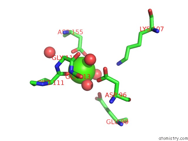
Mono view
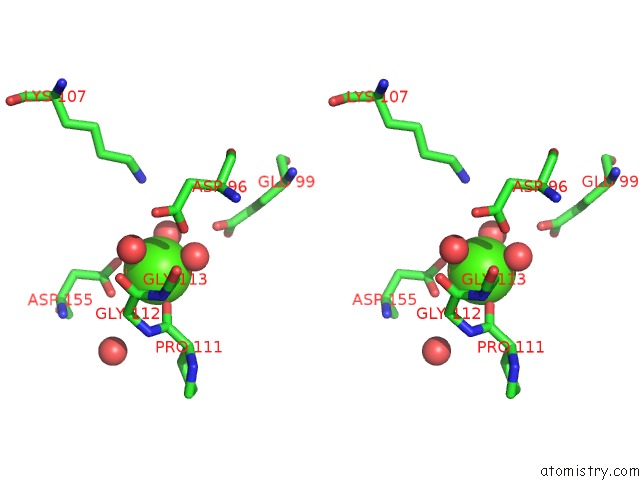
Stereo pair view
Mono view
Stereo pair view
A full contact list of Calcium with other atoms in the Ca binding
site number 1 of Crystal Structure of Catechol 2,3-Dioxygenase within 5.0Å range:
|
Reference:
H.J.Cho,
K.Kim,
S.Y.Sohn,
H.Y.Cho,
K.J.Kim,
M.H.Kim,
D.Kim,
E.Kim,
B.S.Kang.
Substrate-Binding Mechanism of A Type I Extradiol Dioxygenase. J.Biol.Chem. V. 285 34643 2010.
ISSN: ISSN 0021-9258
PubMed: 20810655
DOI: 10.1074/JBC.M110.130310
Page generated: Tue Jul 8 09:07:37 2025
ISSN: ISSN 0021-9258
PubMed: 20810655
DOI: 10.1074/JBC.M110.130310
Last articles
I in 4UR2I in 4UYU
I in 4W6Z
I in 4V1H
I in 4TVD
I in 4U5W
I in 4UJ2
I in 4UE7
I in 4TVC
I in 4TTU