Calcium »
PDB 2xc5-2xon »
2xmo »
Calcium in PDB 2xmo: The Crystal Structure of LMO2642
Protein crystallography data
The structure of The Crystal Structure of LMO2642, PDB code: 2xmo
was solved by
J.H.Jeong,
Y.G.Kim,
with X-Ray Crystallography technique. A brief refinement statistics is given in the table below:
| Resolution Low / High (Å) | 30 / 1.70 |
| Space group | P 1 21 1 |
| Cell size a, b, c (Å), α, β, γ (°) | 62.963, 103.267, 73.765, 90.00, 102.80, 90.00 |
| R / Rfree (%) | 21.18 / 24.25 |
Other elements in 2xmo:
The structure of The Crystal Structure of LMO2642 also contains other interesting chemical elements:
| Manganese | (Mn) | 2 atoms |
| Iron | (Fe) | 2 atoms |
Calcium Binding Sites:
The binding sites of Calcium atom in the The Crystal Structure of LMO2642
(pdb code 2xmo). This binding sites where shown within
5.0 Angstroms radius around Calcium atom.
In total 2 binding sites of Calcium where determined in the The Crystal Structure of LMO2642, PDB code: 2xmo:
Jump to Calcium binding site number: 1; 2;
In total 2 binding sites of Calcium where determined in the The Crystal Structure of LMO2642, PDB code: 2xmo:
Jump to Calcium binding site number: 1; 2;
Calcium binding site 1 out of 2 in 2xmo
Go back to
Calcium binding site 1 out
of 2 in the The Crystal Structure of LMO2642
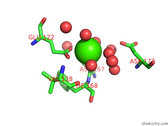
Mono view
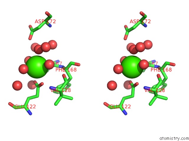
Stereo pair view
Mono view
Stereo pair view
A full contact list of Calcium with other atoms in the Ca binding
site number 1 of The Crystal Structure of LMO2642 within 5.0Å range:
|
Calcium binding site 2 out of 2 in 2xmo
Go back to
Calcium binding site 2 out
of 2 in the The Crystal Structure of LMO2642
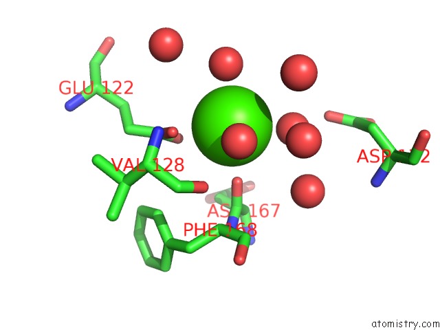
Mono view
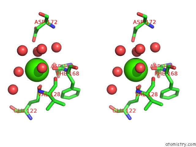
Stereo pair view
Mono view
Stereo pair view
A full contact list of Calcium with other atoms in the Ca binding
site number 2 of The Crystal Structure of LMO2642 within 5.0Å range:
|
Reference:
Y.G.Kim,
J.H.Jeong,
N.C.Ha,
K.J.Kim.
Structural and Functional Analysis of the LMO2642 Cyclic Nucleotide Phosphodiesterase From Listeria Monocytogenes. Proteins V. 79 1205 2011.
ISSN: ISSN 0887-3585
PubMed: 21246635
DOI: 10.1002/PROT.22954
Page generated: Tue Jul 8 09:25:45 2025
ISSN: ISSN 0887-3585
PubMed: 21246635
DOI: 10.1002/PROT.22954
Last articles
K in 3ISFK in 3IGI
K in 3HQP
K in 3IRP
K in 3IQN
K in 3IK4
K in 3IMQ
K in 3IKF
K in 3IIS
K in 3IIN