Calcium »
PDB 2xqg-2y6h »
2xta »
Calcium in PDB 2xta: Crystal Structure of the Suca Domain of Mycobacterium Smegmatis Alpha-Ketoglutarate Decarboxylase in Complex with Acetyl-Coa (Triclinic Form)
Enzymatic activity of Crystal Structure of the Suca Domain of Mycobacterium Smegmatis Alpha-Ketoglutarate Decarboxylase in Complex with Acetyl-Coa (Triclinic Form)
All present enzymatic activity of Crystal Structure of the Suca Domain of Mycobacterium Smegmatis Alpha-Ketoglutarate Decarboxylase in Complex with Acetyl-Coa (Triclinic Form):
4.1.1.71;
4.1.1.71;
Protein crystallography data
The structure of Crystal Structure of the Suca Domain of Mycobacterium Smegmatis Alpha-Ketoglutarate Decarboxylase in Complex with Acetyl-Coa (Triclinic Form), PDB code: 2xta
was solved by
T.Wagner,
M.Bellinzoni,
A.M.Wehenkel,
H.M.O'hare,
P.M.Alzari,
with X-Ray Crystallography technique. A brief refinement statistics is given in the table below:
| Resolution Low / High (Å) | 78.11 / 2.20 |
| Space group | P 1 |
| Cell size a, b, c (Å), α, β, γ (°) | 80.550, 83.580, 160.070, 99.59, 98.94, 100.68 |
| R / Rfree (%) | 18.8 / 21.36 |
Other elements in 2xta:
The structure of Crystal Structure of the Suca Domain of Mycobacterium Smegmatis Alpha-Ketoglutarate Decarboxylase in Complex with Acetyl-Coa (Triclinic Form) also contains other interesting chemical elements:
| Magnesium | (Mg) | 4 atoms |
Calcium Binding Sites:
The binding sites of Calcium atom in the Crystal Structure of the Suca Domain of Mycobacterium Smegmatis Alpha-Ketoglutarate Decarboxylase in Complex with Acetyl-Coa (Triclinic Form)
(pdb code 2xta). This binding sites where shown within
5.0 Angstroms radius around Calcium atom.
In total 4 binding sites of Calcium where determined in the Crystal Structure of the Suca Domain of Mycobacterium Smegmatis Alpha-Ketoglutarate Decarboxylase in Complex with Acetyl-Coa (Triclinic Form), PDB code: 2xta:
Jump to Calcium binding site number: 1; 2; 3; 4;
In total 4 binding sites of Calcium where determined in the Crystal Structure of the Suca Domain of Mycobacterium Smegmatis Alpha-Ketoglutarate Decarboxylase in Complex with Acetyl-Coa (Triclinic Form), PDB code: 2xta:
Jump to Calcium binding site number: 1; 2; 3; 4;
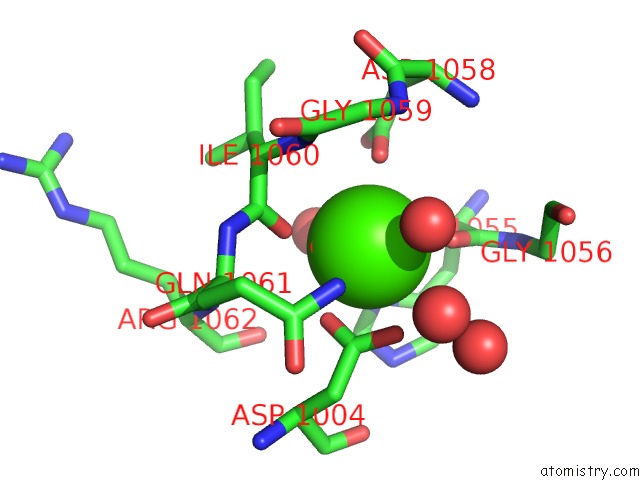
Calcium binding site 1 out of 4 in 2xta
Go back to
Calcium binding site 1 out
of 4 in the Crystal Structure of the Suca Domain of Mycobacterium Smegmatis Alpha-Ketoglutarate Decarboxylase in Complex with Acetyl-Coa (Triclinic Form)
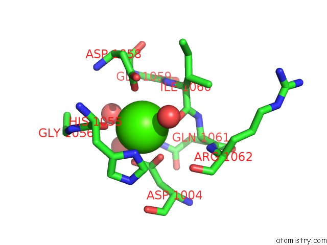
Mono view
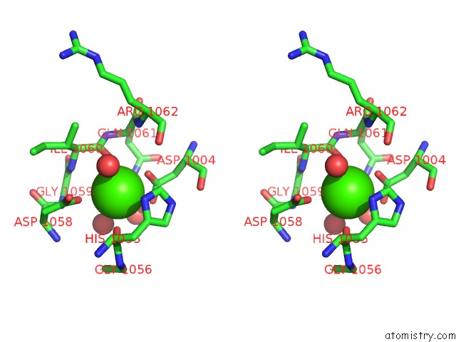
Stereo pair view
Mono view
Stereo pair view
A full contact list of Calcium with other atoms in the Ca binding
site number 1 of Crystal Structure of the Suca Domain of Mycobacterium Smegmatis Alpha-Ketoglutarate Decarboxylase in Complex with Acetyl-Coa (Triclinic Form) within 5.0Å range:
|
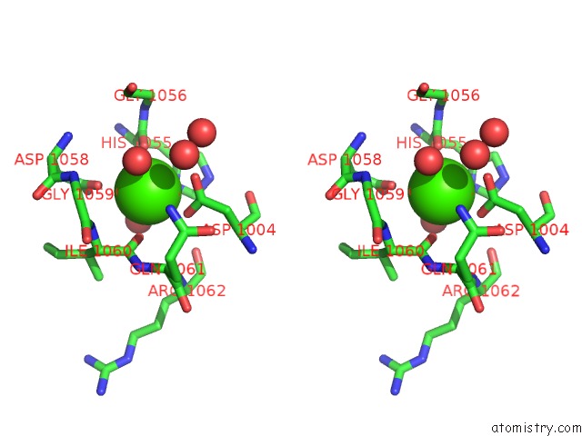
Calcium binding site 2 out of 4 in 2xta
Go back to
Calcium binding site 2 out
of 4 in the Crystal Structure of the Suca Domain of Mycobacterium Smegmatis Alpha-Ketoglutarate Decarboxylase in Complex with Acetyl-Coa (Triclinic Form)
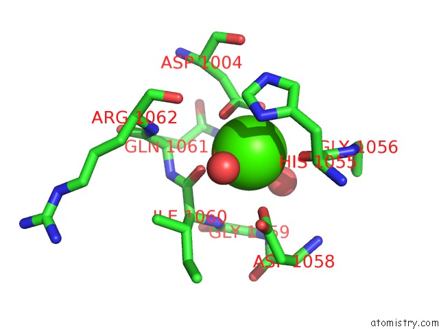
Mono view
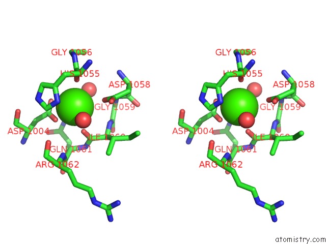
Stereo pair view
Mono view
Stereo pair view
A full contact list of Calcium with other atoms in the Ca binding
site number 2 of Crystal Structure of the Suca Domain of Mycobacterium Smegmatis Alpha-Ketoglutarate Decarboxylase in Complex with Acetyl-Coa (Triclinic Form) within 5.0Å range:
|
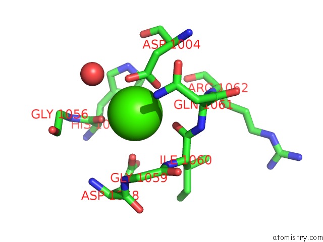
Calcium binding site 3 out of 4 in 2xta
Go back to
Calcium binding site 3 out
of 4 in the Crystal Structure of the Suca Domain of Mycobacterium Smegmatis Alpha-Ketoglutarate Decarboxylase in Complex with Acetyl-Coa (Triclinic Form)
Mono view
Stereo pair view
Mono view
Stereo pair view
A full contact list of Calcium with other atoms in the Ca binding
site number 3 of Crystal Structure of the Suca Domain of Mycobacterium Smegmatis Alpha-Ketoglutarate Decarboxylase in Complex with Acetyl-Coa (Triclinic Form) within 5.0Å range:
|
Calcium binding site 4 out of 4 in 2xta
Go back to
Calcium binding site 4 out
of 4 in the Crystal Structure of the Suca Domain of Mycobacterium Smegmatis Alpha-Ketoglutarate Decarboxylase in Complex with Acetyl-Coa (Triclinic Form)
Mono view
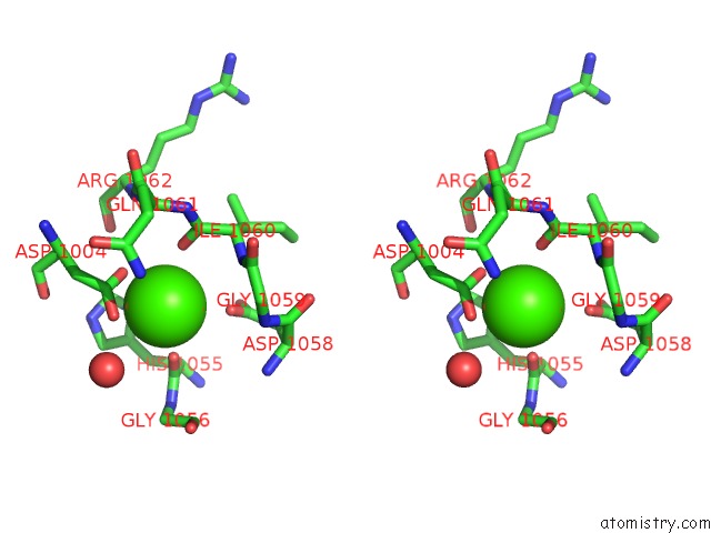
Stereo pair view
Mono view
Stereo pair view
A full contact list of Calcium with other atoms in the Ca binding
site number 4 of Crystal Structure of the Suca Domain of Mycobacterium Smegmatis Alpha-Ketoglutarate Decarboxylase in Complex with Acetyl-Coa (Triclinic Form) within 5.0Å range:
|
Reference:
T.Wagner,
M.Bellinzoni,
A.M.Wehenkel,
H.M.O'hare,
P.M.Alzari.
Functional Plasticity and Allosteric Regulation of Alpha-Ketoglutarate Decarboxylase in Central Mycobacterial Metabolism. Chem.Biol. V. 18 1011 2011.
ISSN: ISSN 1074-5521
PubMed: 21867916
DOI: 10.1016/J.CHEMBIOL.2011.06.004
Page generated: Tue Jul 8 09:28:49 2025
ISSN: ISSN 1074-5521
PubMed: 21867916
DOI: 10.1016/J.CHEMBIOL.2011.06.004
Last articles
K in 4BH5K in 4B3T
K in 4BG8
K in 4BG9
K in 4B5R
K in 4BEW
K in 4BE6
K in 4B7V
K in 4B3M
K in 4B3S