Calcium »
PDB 2xqg-2y6h »
2y6g »
Calcium in PDB 2y6g: Cellopentaose Binding Mutated (X-2 L110F) CBM4-2 Carbohydrate Binding Module From A Thermostable Rhodothermus Marinus Xylanase
Enzymatic activity of Cellopentaose Binding Mutated (X-2 L110F) CBM4-2 Carbohydrate Binding Module From A Thermostable Rhodothermus Marinus Xylanase
All present enzymatic activity of Cellopentaose Binding Mutated (X-2 L110F) CBM4-2 Carbohydrate Binding Module From A Thermostable Rhodothermus Marinus Xylanase:
3.2.1.8;
3.2.1.8;
Protein crystallography data
The structure of Cellopentaose Binding Mutated (X-2 L110F) CBM4-2 Carbohydrate Binding Module From A Thermostable Rhodothermus Marinus Xylanase, PDB code: 2y6g
was solved by
L.Von Schantz,
M.Hakansson,
D.T.Logan,
B.Walse,
J.Osterlin,
E.Nordberg-Karlsson,
M.Ohlin,
with X-Ray Crystallography technique. A brief refinement statistics is given in the table below:
| Resolution Low / High (Å) | 30.00 / 1.30 |
| Space group | P 21 21 21 |
| Cell size a, b, c (Å), α, β, γ (°) | 48.560, 49.720, 62.520, 90.00, 90.00, 90.00 |
| R / Rfree (%) | 13.2 / 18.3 |
Calcium Binding Sites:
The binding sites of Calcium atom in the Cellopentaose Binding Mutated (X-2 L110F) CBM4-2 Carbohydrate Binding Module From A Thermostable Rhodothermus Marinus Xylanase
(pdb code 2y6g). This binding sites where shown within
5.0 Angstroms radius around Calcium atom.
In total 2 binding sites of Calcium where determined in the Cellopentaose Binding Mutated (X-2 L110F) CBM4-2 Carbohydrate Binding Module From A Thermostable Rhodothermus Marinus Xylanase, PDB code: 2y6g:
Jump to Calcium binding site number: 1; 2;
In total 2 binding sites of Calcium where determined in the Cellopentaose Binding Mutated (X-2 L110F) CBM4-2 Carbohydrate Binding Module From A Thermostable Rhodothermus Marinus Xylanase, PDB code: 2y6g:
Jump to Calcium binding site number: 1; 2;
Calcium binding site 1 out of 2 in 2y6g
Go back to
Calcium binding site 1 out
of 2 in the Cellopentaose Binding Mutated (X-2 L110F) CBM4-2 Carbohydrate Binding Module From A Thermostable Rhodothermus Marinus Xylanase
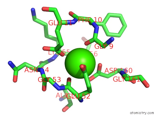
Mono view
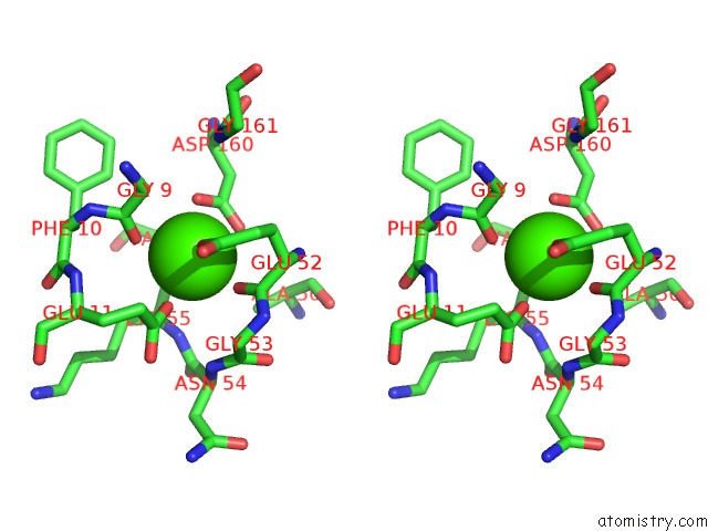
Stereo pair view
Mono view
Stereo pair view
A full contact list of Calcium with other atoms in the Ca binding
site number 1 of Cellopentaose Binding Mutated (X-2 L110F) CBM4-2 Carbohydrate Binding Module From A Thermostable Rhodothermus Marinus Xylanase within 5.0Å range:
|
Calcium binding site 2 out of 2 in 2y6g
Go back to
Calcium binding site 2 out
of 2 in the Cellopentaose Binding Mutated (X-2 L110F) CBM4-2 Carbohydrate Binding Module From A Thermostable Rhodothermus Marinus Xylanase
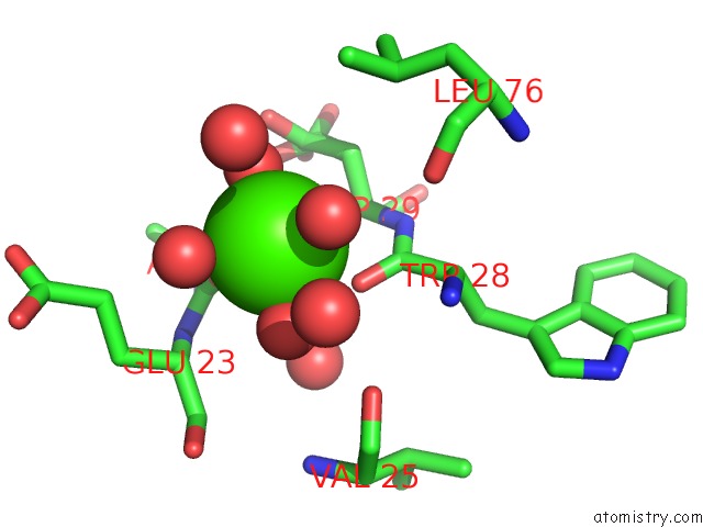
Mono view
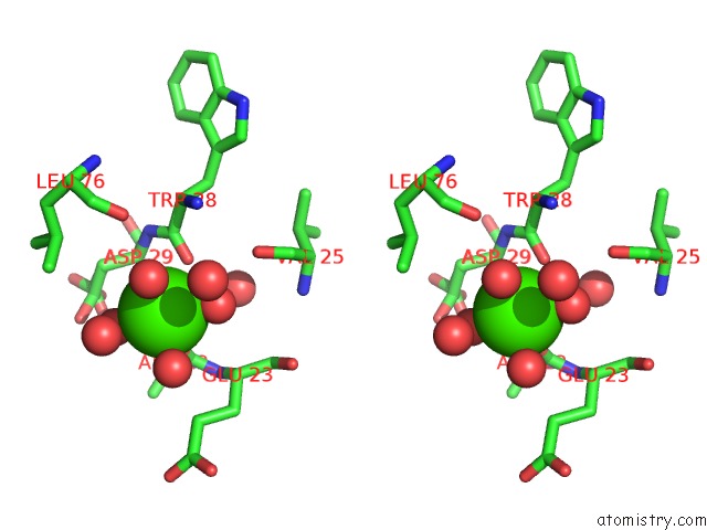
Stereo pair view
Mono view
Stereo pair view
A full contact list of Calcium with other atoms in the Ca binding
site number 2 of Cellopentaose Binding Mutated (X-2 L110F) CBM4-2 Carbohydrate Binding Module From A Thermostable Rhodothermus Marinus Xylanase within 5.0Å range:
|
Reference:
L.Von Schantz,
M.Hakansson,
D.T.Logan,
B.Walse,
J.Osterlin,
E.Nordberg-Karlsson,
M.Ohlin.
Structural Basis For Carbohydrate-Binding Specificity--A Comparative Assessment of Two Engineered Carbohydrate-Binding Modules. Glycobiology V. 22 948 2012.
ISSN: ESSN 1460-2423
PubMed: 22434778
DOI: 10.1093/GLYCOB/CWS063
Page generated: Tue Jul 8 09:33:58 2025
ISSN: ESSN 1460-2423
PubMed: 22434778
DOI: 10.1093/GLYCOB/CWS063
Last articles
K in 8YZ7K in 8XW8
K in 8XW9
K in 8XTS
K in 8XTR
K in 8XW7
K in 8XW6
K in 8XTQ
K in 8XMI
K in 8XTP