Calcium »
PDB 2yi1-2z8s »
2yic »
Calcium in PDB 2yic: Crystal Structure of the Suca Domain of Mycobacterium Smegmatis Alpha-Ketoglutarate Decarboxylase (Triclinic Form)
Enzymatic activity of Crystal Structure of the Suca Domain of Mycobacterium Smegmatis Alpha-Ketoglutarate Decarboxylase (Triclinic Form)
All present enzymatic activity of Crystal Structure of the Suca Domain of Mycobacterium Smegmatis Alpha-Ketoglutarate Decarboxylase (Triclinic Form):
4.1.1.71;
4.1.1.71;
Protein crystallography data
The structure of Crystal Structure of the Suca Domain of Mycobacterium Smegmatis Alpha-Ketoglutarate Decarboxylase (Triclinic Form), PDB code: 2yic
was solved by
T.Wagner,
M.Bellinzoni,
A.M.Wehenkel,
H.M.O'hare,
P.M.Alzari,
with X-Ray Crystallography technique. A brief refinement statistics is given in the table below:
| Resolution Low / High (Å) | 39.20 / 1.96 |
| Space group | P 1 |
| Cell size a, b, c (Å), α, β, γ (°) | 79.539, 83.244, 158.610, 99.48, 99.06, 101.25 |
| R / Rfree (%) | 18.84 / 21.08 |
Other elements in 2yic:
The structure of Crystal Structure of the Suca Domain of Mycobacterium Smegmatis Alpha-Ketoglutarate Decarboxylase (Triclinic Form) also contains other interesting chemical elements:
| Magnesium | (Mg) | 4 atoms |
Calcium Binding Sites:
The binding sites of Calcium atom in the Crystal Structure of the Suca Domain of Mycobacterium Smegmatis Alpha-Ketoglutarate Decarboxylase (Triclinic Form)
(pdb code 2yic). This binding sites where shown within
5.0 Angstroms radius around Calcium atom.
In total 4 binding sites of Calcium where determined in the Crystal Structure of the Suca Domain of Mycobacterium Smegmatis Alpha-Ketoglutarate Decarboxylase (Triclinic Form), PDB code: 2yic:
Jump to Calcium binding site number: 1; 2; 3; 4;
In total 4 binding sites of Calcium where determined in the Crystal Structure of the Suca Domain of Mycobacterium Smegmatis Alpha-Ketoglutarate Decarboxylase (Triclinic Form), PDB code: 2yic:
Jump to Calcium binding site number: 1; 2; 3; 4;
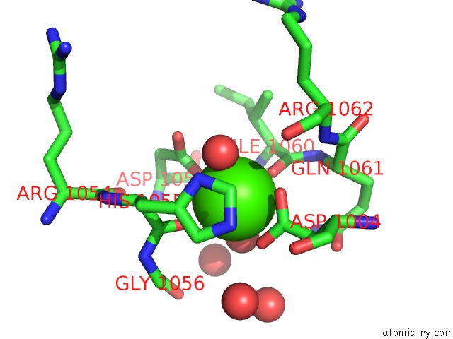
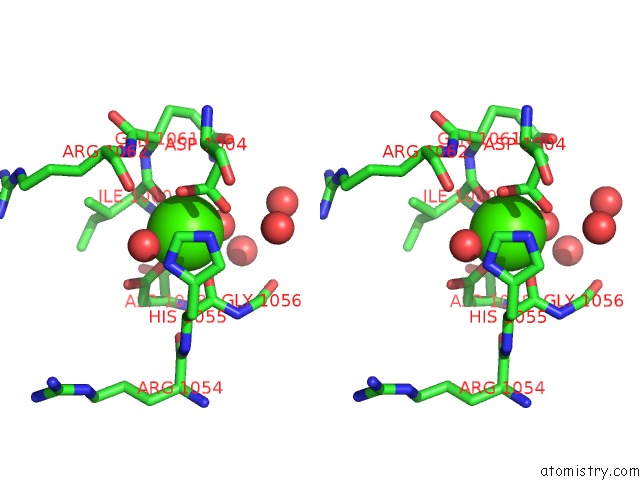
Calcium binding site 1 out of 4 in 2yic
Go back to
Calcium binding site 1 out
of 4 in the Crystal Structure of the Suca Domain of Mycobacterium Smegmatis Alpha-Ketoglutarate Decarboxylase (Triclinic Form)
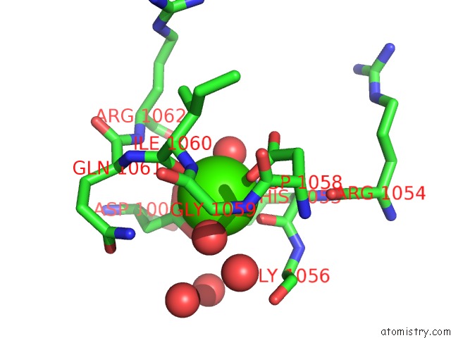
Mono view
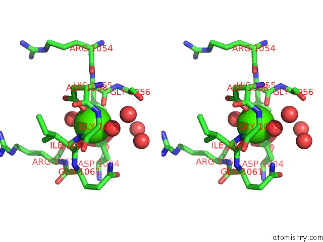
Stereo pair view
Mono view
Stereo pair view
A full contact list of Calcium with other atoms in the Ca binding
site number 1 of Crystal Structure of the Suca Domain of Mycobacterium Smegmatis Alpha-Ketoglutarate Decarboxylase (Triclinic Form) within 5.0Å range:
|
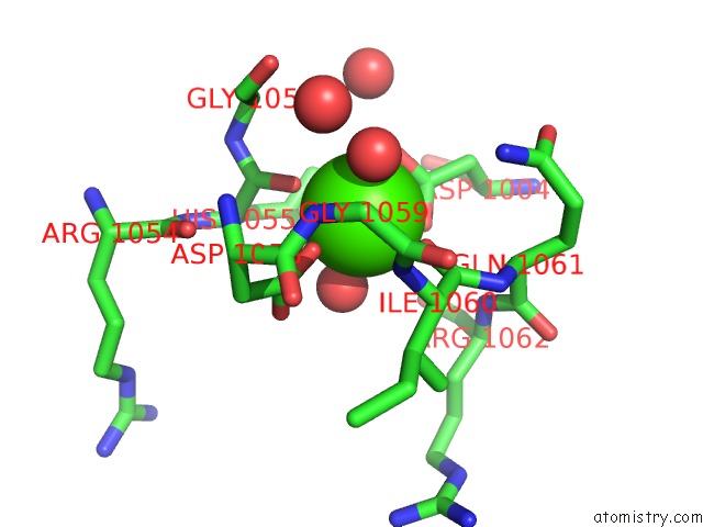
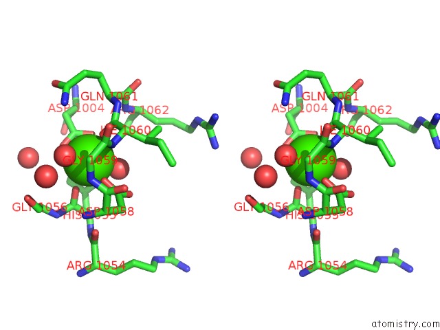
Calcium binding site 2 out of 4 in 2yic
Go back to
Calcium binding site 2 out
of 4 in the Crystal Structure of the Suca Domain of Mycobacterium Smegmatis Alpha-Ketoglutarate Decarboxylase (Triclinic Form)
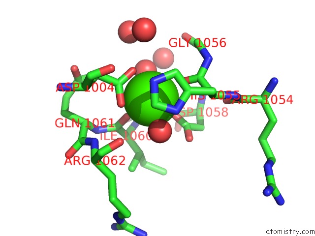
Mono view
Stereo pair view
Mono view
Stereo pair view
A full contact list of Calcium with other atoms in the Ca binding
site number 2 of Crystal Structure of the Suca Domain of Mycobacterium Smegmatis Alpha-Ketoglutarate Decarboxylase (Triclinic Form) within 5.0Å range:
|
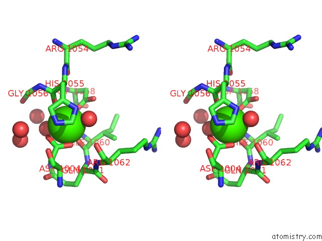
Calcium binding site 3 out of 4 in 2yic
Go back to
Calcium binding site 3 out
of 4 in the Crystal Structure of the Suca Domain of Mycobacterium Smegmatis Alpha-Ketoglutarate Decarboxylase (Triclinic Form)
Mono view
Stereo pair view
Mono view
Stereo pair view
A full contact list of Calcium with other atoms in the Ca binding
site number 3 of Crystal Structure of the Suca Domain of Mycobacterium Smegmatis Alpha-Ketoglutarate Decarboxylase (Triclinic Form) within 5.0Å range:
|
Calcium binding site 4 out of 4 in 2yic
Go back to
Calcium binding site 4 out
of 4 in the Crystal Structure of the Suca Domain of Mycobacterium Smegmatis Alpha-Ketoglutarate Decarboxylase (Triclinic Form)
Mono view
Stereo pair view
Mono view
Stereo pair view
A full contact list of Calcium with other atoms in the Ca binding
site number 4 of Crystal Structure of the Suca Domain of Mycobacterium Smegmatis Alpha-Ketoglutarate Decarboxylase (Triclinic Form) within 5.0Å range:
|
Reference:
T.Wagner,
M.Bellinzoni,
A.M.Wehenkel,
H.M.O'hare,
P.M.Alzari.
Functional Plasticity and Allosteric Regulation of Alpha-Ketoglutarate Decarboxylase in Central Mycobacterial Metabolism. Chem.Biol. V. 18 1011 2011.
ISSN: ISSN 1074-5521
PubMed: 21867916
DOI: 10.1016/J.CHEMBIOL.2011.06.004
Page generated: Tue Jul 8 09:40:28 2025
ISSN: ISSN 1074-5521
PubMed: 21867916
DOI: 10.1016/J.CHEMBIOL.2011.06.004
Last articles
Mg in 3TWAMg in 3TW0
Mg in 3TW7
Mg in 3TVY
Mg in 3TVX
Mg in 3TVD
Mg in 3TTZ
Mg in 3TVK
Mg in 3TVB
Mg in 3TVA