Calcium »
PDB 2z8x-2zp4 »
2zid »
Calcium in PDB 2zid: Crystal Structure of Dextran Glucosidase E236Q Complex with Isomaltotriose
Enzymatic activity of Crystal Structure of Dextran Glucosidase E236Q Complex with Isomaltotriose
All present enzymatic activity of Crystal Structure of Dextran Glucosidase E236Q Complex with Isomaltotriose:
3.2.1.70;
3.2.1.70;
Protein crystallography data
The structure of Crystal Structure of Dextran Glucosidase E236Q Complex with Isomaltotriose, PDB code: 2zid
was solved by
H.Hondoh,
W.Saburi,
H.Mori,
M.Okuyama,
T.Nakada,
Y.Matsuura,
A.Kimura,
with X-Ray Crystallography technique. A brief refinement statistics is given in the table below:
| Resolution Low / High (Å) | 10.00 / 2.20 |
| Space group | P 21 21 21 |
| Cell size a, b, c (Å), α, β, γ (°) | 72.490, 82.530, 104.300, 90.00, 90.00, 90.00 |
| R / Rfree (%) | 18.4 / 22.4 |
Calcium Binding Sites:
The binding sites of Calcium atom in the Crystal Structure of Dextran Glucosidase E236Q Complex with Isomaltotriose
(pdb code 2zid). This binding sites where shown within
5.0 Angstroms radius around Calcium atom.
In total 3 binding sites of Calcium where determined in the Crystal Structure of Dextran Glucosidase E236Q Complex with Isomaltotriose, PDB code: 2zid:
Jump to Calcium binding site number: 1; 2; 3;
In total 3 binding sites of Calcium where determined in the Crystal Structure of Dextran Glucosidase E236Q Complex with Isomaltotriose, PDB code: 2zid:
Jump to Calcium binding site number: 1; 2; 3;
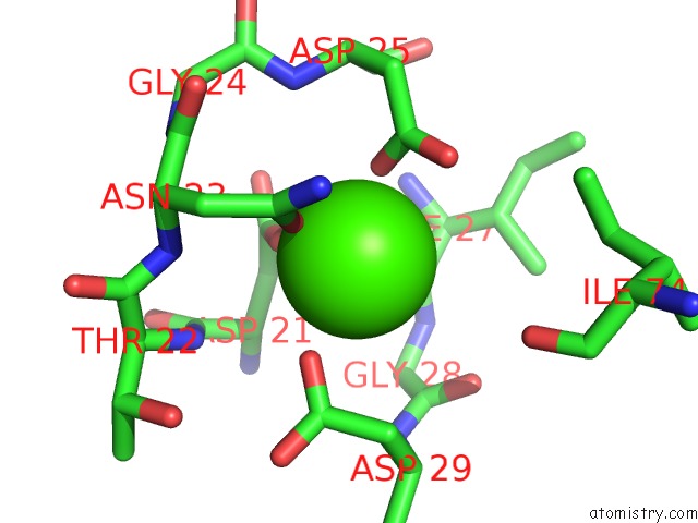
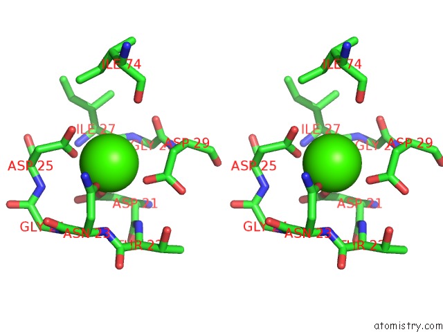
Calcium binding site 1 out of 3 in 2zid
Go back to
Calcium binding site 1 out
of 3 in the Crystal Structure of Dextran Glucosidase E236Q Complex with Isomaltotriose
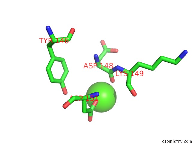
Mono view
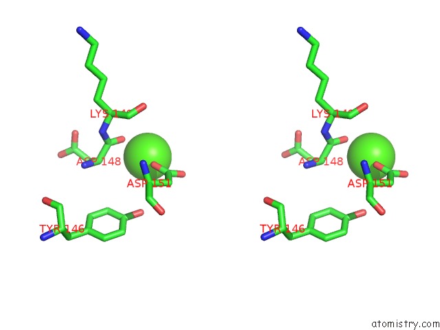
Stereo pair view
Mono view
Stereo pair view
A full contact list of Calcium with other atoms in the Ca binding
site number 1 of Crystal Structure of Dextran Glucosidase E236Q Complex with Isomaltotriose within 5.0Å range:
|
Calcium binding site 2 out of 3 in 2zid
Go back to
Calcium binding site 2 out
of 3 in the Crystal Structure of Dextran Glucosidase E236Q Complex with Isomaltotriose
Mono view
Stereo pair view
Mono view
Stereo pair view
A full contact list of Calcium with other atoms in the Ca binding
site number 2 of Crystal Structure of Dextran Glucosidase E236Q Complex with Isomaltotriose within 5.0Å range:
|
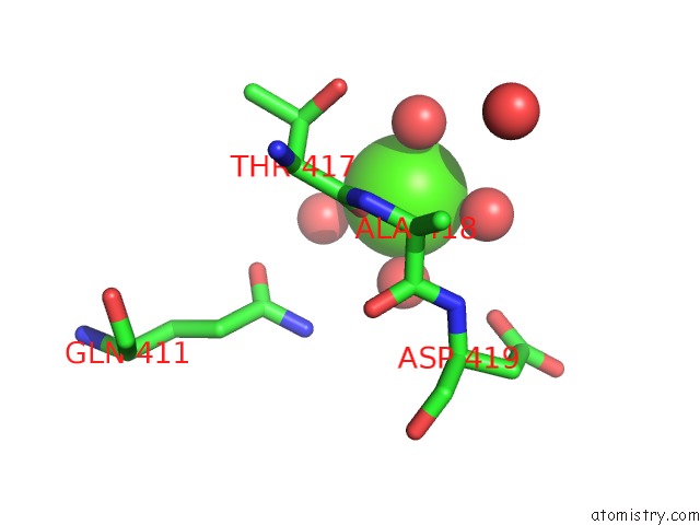
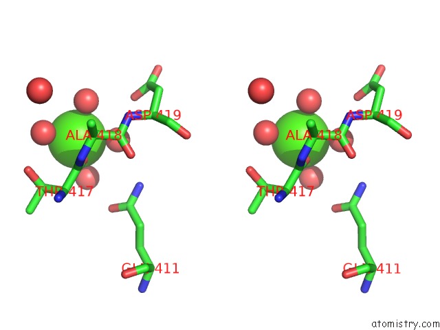
Calcium binding site 3 out of 3 in 2zid
Go back to
Calcium binding site 3 out
of 3 in the Crystal Structure of Dextran Glucosidase E236Q Complex with Isomaltotriose
Mono view
Stereo pair view
Mono view
Stereo pair view
A full contact list of Calcium with other atoms in the Ca binding
site number 3 of Crystal Structure of Dextran Glucosidase E236Q Complex with Isomaltotriose within 5.0Å range:
|
Reference:
H.Hondoh,
W.Saburi,
H.Mori,
M.Okuyama,
T.Nakada,
Y.Matsuura,
A.Kimura.
Substrate Recognition Mechanism of Alpha-1,6-Glucosidic Linkage Hydrolyzing Enzyme, Dextran Glucosidase From Streptococcus Mutans. J.Mol.Biol. V. 378 911 2008.
ISSN: ISSN 0022-2836
PubMed: 18395742
DOI: 10.1016/J.JMB.2008.03.016
Page generated: Tue Jul 8 10:02:37 2025
ISSN: ISSN 0022-2836
PubMed: 18395742
DOI: 10.1016/J.JMB.2008.03.016
Last articles
Fe in 2YXOFe in 2YRS
Fe in 2YXC
Fe in 2YNM
Fe in 2YVJ
Fe in 2YP1
Fe in 2YU2
Fe in 2YU1
Fe in 2YQB
Fe in 2YOO