Calcium »
PDB 2zp5-3a4h »
3a13 »
Calcium in PDB 3a13: Crystal Structure of Type III Rubisco SP4 Mutant Complexed with 2-Cabp and Activated with Ca
Enzymatic activity of Crystal Structure of Type III Rubisco SP4 Mutant Complexed with 2-Cabp and Activated with Ca
All present enzymatic activity of Crystal Structure of Type III Rubisco SP4 Mutant Complexed with 2-Cabp and Activated with Ca:
4.1.1.39;
4.1.1.39;
Protein crystallography data
The structure of Crystal Structure of Type III Rubisco SP4 Mutant Complexed with 2-Cabp and Activated with Ca, PDB code: 3a13
was solved by
Y.Nishitani,
M.Fujihashi,
T.Doi,
S.Yoshida,
H.Atomi,
T.Imanaka,
K.Miki,
with X-Ray Crystallography technique. A brief refinement statistics is given in the table below:
| Resolution Low / High (Å) | 42.22 / 2.34 |
| Space group | P 21 21 2 |
| Cell size a, b, c (Å), α, β, γ (°) | 173.678, 247.090, 144.940, 90.00, 90.00, 90.00 |
| R / Rfree (%) | 20.5 / 25 |
Other elements in 3a13:
The structure of Crystal Structure of Type III Rubisco SP4 Mutant Complexed with 2-Cabp and Activated with Ca also contains other interesting chemical elements:
| Magnesium | (Mg) | 8 atoms |
Calcium Binding Sites:
The binding sites of Calcium atom in the Crystal Structure of Type III Rubisco SP4 Mutant Complexed with 2-Cabp and Activated with Ca
(pdb code 3a13). This binding sites where shown within
5.0 Angstroms radius around Calcium atom.
In total 2 binding sites of Calcium where determined in the Crystal Structure of Type III Rubisco SP4 Mutant Complexed with 2-Cabp and Activated with Ca, PDB code: 3a13:
Jump to Calcium binding site number: 1; 2;
In total 2 binding sites of Calcium where determined in the Crystal Structure of Type III Rubisco SP4 Mutant Complexed with 2-Cabp and Activated with Ca, PDB code: 3a13:
Jump to Calcium binding site number: 1; 2;
Calcium binding site 1 out of 2 in 3a13
Go back to
Calcium binding site 1 out
of 2 in the Crystal Structure of Type III Rubisco SP4 Mutant Complexed with 2-Cabp and Activated with Ca
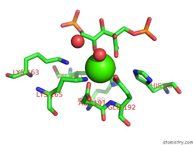
Mono view
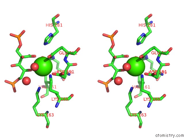
Stereo pair view
Mono view
Stereo pair view
A full contact list of Calcium with other atoms in the Ca binding
site number 1 of Crystal Structure of Type III Rubisco SP4 Mutant Complexed with 2-Cabp and Activated with Ca within 5.0Å range:
|
Calcium binding site 2 out of 2 in 3a13
Go back to
Calcium binding site 2 out
of 2 in the Crystal Structure of Type III Rubisco SP4 Mutant Complexed with 2-Cabp and Activated with Ca
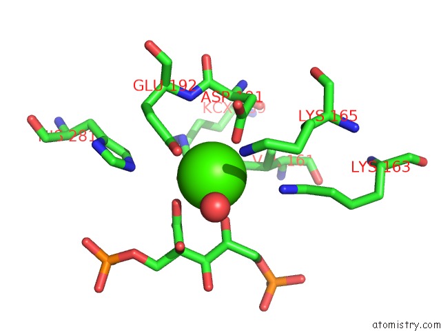
Mono view
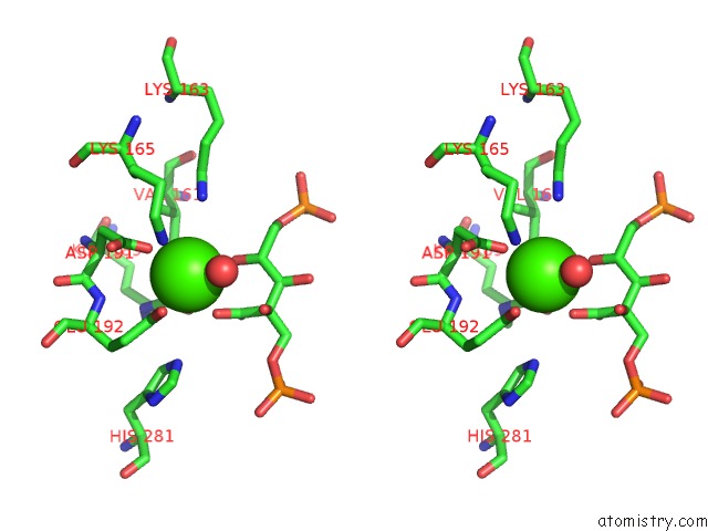
Stereo pair view
Mono view
Stereo pair view
A full contact list of Calcium with other atoms in the Ca binding
site number 2 of Crystal Structure of Type III Rubisco SP4 Mutant Complexed with 2-Cabp and Activated with Ca within 5.0Å range:
|
Reference:
Y.Nishitani,
M.Fujihashi,
T.Doi,
S.Yoshida,
H.Atomi,
T.Imanaka,
K.Miki.
Sturcture-Based Optimization of A Type III Rubisco From A Hyperthermophile To Be Published.
Page generated: Tue Jul 8 10:21:23 2025
Last articles
Mg in 5SCEMg in 5SCF
Mg in 5SCC
Mg in 5SCD
Mg in 5SC9
Mg in 5SCB
Mg in 5SC8
Mg in 5SCA
Mg in 5SBE
Mg in 5SBD