Calcium »
PDB 3b1u-3biw »
3b7y »
Calcium in PDB 3b7y: Crystal Structure of the C2 Domain of the E3 Ubiquitin- Protein Ligase NEDD4
Protein crystallography data
The structure of Crystal Structure of the C2 Domain of the E3 Ubiquitin- Protein Ligase NEDD4, PDB code: 3b7y
was solved by
J.R.Walker,
M.Ruzanov,
C.Butler-Cole,
J.Weigelt,
C.H.Arrowsmith,
A.M.Edwards,
A.Bochkarev,
S.Dhe-Paganon,
Structural Genomicsconsortium (Sgc),
with X-Ray Crystallography technique. A brief refinement statistics is given in the table below:
| Resolution Low / High (Å) | 27.23 / 1.80 |
| Space group | P 1 |
| Cell size a, b, c (Å), α, β, γ (°) | 38.778, 47.997, 50.786, 108.89, 111.11, 100.86 |
| R / Rfree (%) | 16.9 / 20.9 |
Calcium Binding Sites:
The binding sites of Calcium atom in the Crystal Structure of the C2 Domain of the E3 Ubiquitin- Protein Ligase NEDD4
(pdb code 3b7y). This binding sites where shown within
5.0 Angstroms radius around Calcium atom.
In total 2 binding sites of Calcium where determined in the Crystal Structure of the C2 Domain of the E3 Ubiquitin- Protein Ligase NEDD4, PDB code: 3b7y:
Jump to Calcium binding site number: 1; 2;
In total 2 binding sites of Calcium where determined in the Crystal Structure of the C2 Domain of the E3 Ubiquitin- Protein Ligase NEDD4, PDB code: 3b7y:
Jump to Calcium binding site number: 1; 2;
Calcium binding site 1 out of 2 in 3b7y
Go back to
Calcium binding site 1 out
of 2 in the Crystal Structure of the C2 Domain of the E3 Ubiquitin- Protein Ligase NEDD4
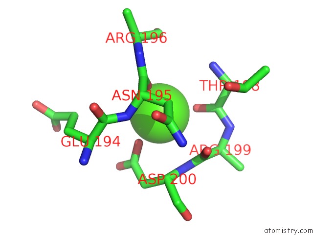
Mono view
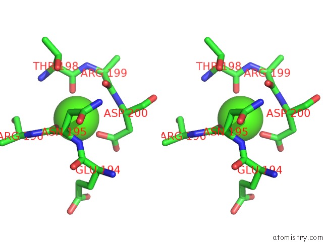
Stereo pair view
Mono view
Stereo pair view
A full contact list of Calcium with other atoms in the Ca binding
site number 1 of Crystal Structure of the C2 Domain of the E3 Ubiquitin- Protein Ligase NEDD4 within 5.0Å range:
|
Calcium binding site 2 out of 2 in 3b7y
Go back to
Calcium binding site 2 out
of 2 in the Crystal Structure of the C2 Domain of the E3 Ubiquitin- Protein Ligase NEDD4
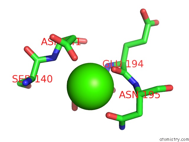
Mono view
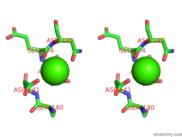
Stereo pair view
Mono view
Stereo pair view
A full contact list of Calcium with other atoms in the Ca binding
site number 2 of Crystal Structure of the C2 Domain of the E3 Ubiquitin- Protein Ligase NEDD4 within 5.0Å range:
|
Reference:
J.R.Walker,
M.Ruzanov,
C.Butler-Cole,
J.Weigelt,
C.H.Arrowsmith,
A.M.Edwards,
A.Bochkarev,
S.Dhe-Paganon.
C2 Domain of the Human E3 Ubiquitin-Protein Ligase NEDD4. To Be Published.
Page generated: Tue Jul 8 11:02:58 2025
Last articles
Mg in 6JNXMg in 6JMG
Mg in 6JNL
Mg in 6JLV
Mg in 6JLN
Mg in 6JLL
Mg in 6JLK
Mg in 6JLM
Mg in 6JLJ
Mg in 6JIL