Calcium »
PDB 3bje-3bxk »
3bje »
Calcium in PDB 3bje: Crystal Structure of Trypanosoma Brucei Nucleoside Phosphorylase Shows Uridine Phosphorylase Activity
Protein crystallography data
The structure of Crystal Structure of Trypanosoma Brucei Nucleoside Phosphorylase Shows Uridine Phosphorylase Activity, PDB code: 3bje
was solved by
E.T.Larson,
E.A.Merritt,
Structural Genomics Of Pathogenic Protozoaconsortium (Sgpp),
with X-Ray Crystallography technique. A brief refinement statistics is given in the table below:
| Resolution Low / High (Å) | 35.38 / 1.44 |
| Space group | P 1 21 1 |
| Cell size a, b, c (Å), α, β, γ (°) | 63.015, 95.387, 63.483, 90.00, 105.91, 90.00 |
| R / Rfree (%) | 15.5 / 18.4 |
Calcium Binding Sites:
The binding sites of Calcium atom in the Crystal Structure of Trypanosoma Brucei Nucleoside Phosphorylase Shows Uridine Phosphorylase Activity
(pdb code 3bje). This binding sites where shown within
5.0 Angstroms radius around Calcium atom.
In total only one binding site of Calcium was determined in the Crystal Structure of Trypanosoma Brucei Nucleoside Phosphorylase Shows Uridine Phosphorylase Activity, PDB code: 3bje:
In total only one binding site of Calcium was determined in the Crystal Structure of Trypanosoma Brucei Nucleoside Phosphorylase Shows Uridine Phosphorylase Activity, PDB code: 3bje:
Calcium binding site 1 out of 1 in 3bje
Go back to
Calcium binding site 1 out
of 1 in the Crystal Structure of Trypanosoma Brucei Nucleoside Phosphorylase Shows Uridine Phosphorylase Activity
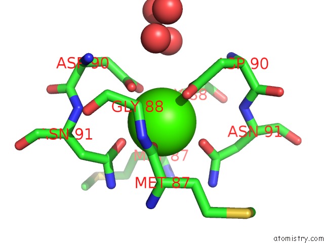
Mono view
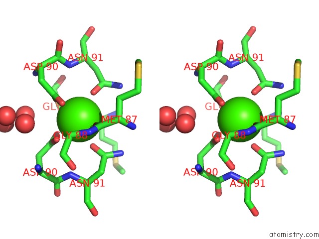
Stereo pair view
Mono view
Stereo pair view
A full contact list of Calcium with other atoms in the Ca binding
site number 1 of Crystal Structure of Trypanosoma Brucei Nucleoside Phosphorylase Shows Uridine Phosphorylase Activity within 5.0Å range:
|
Reference:
E.T.Larson,
D.G.Mudeppa,
J.R.Gillespie,
N.Mueller,
A.J.Napuli,
J.A.Arif,
J.Ross,
T.L.Arakaki,
A.Lauricella,
G.Detitta,
J.Luft,
F.Zucker,
C.L.Verlinde,
E.Fan,
W.C.Van Voorhis,
F.S.Buckner,
P.K.Rathod,
W.G.Hol,
E.A.Merritt.
The Crystal Structure and Activity of A Putative Trypanosomal Nucleoside Phosphorylase Reveal It to Be A Homodimeric Uridine Phosphorylase J.Mol.Biol. V. 396 1244 2010.
ISSN: ISSN 0022-2836
PubMed: 20070944
DOI: 10.1016/J.JMB.2010.01.013
Page generated: Tue Jul 8 11:09:26 2025
ISSN: ISSN 0022-2836
PubMed: 20070944
DOI: 10.1016/J.JMB.2010.01.013
Last articles
Mg in 8H53Mg in 8H68
Mg in 8H5Y
Mg in 8H4P
Mg in 8H5M
Mg in 8H4G
Mg in 8H4F
Mg in 8H4D
Mg in 8H4E
Mg in 8H4C