Calcium »
PDB 3bje-3bxk »
3bs6 »
Calcium in PDB 3bs6: 1.8 Angstrom Crystal Structure of the Periplasmic Domain of the Membrane Insertase Yidc
Protein crystallography data
The structure of 1.8 Angstrom Crystal Structure of the Periplasmic Domain of the Membrane Insertase Yidc, PDB code: 3bs6
was solved by
S.Ravaud,
I.Sinning,
with X-Ray Crystallography technique. A brief refinement statistics is given in the table below:
| Resolution Low / High (Å) | 38.90 / 1.80 |
| Space group | C 1 2 1 |
| Cell size a, b, c (Å), α, β, γ (°) | 161.150, 55.640, 63.330, 90.00, 101.12, 90.00 |
| R / Rfree (%) | 17.8 / 21.3 |
Calcium Binding Sites:
The binding sites of Calcium atom in the 1.8 Angstrom Crystal Structure of the Periplasmic Domain of the Membrane Insertase Yidc
(pdb code 3bs6). This binding sites where shown within
5.0 Angstroms radius around Calcium atom.
In total 5 binding sites of Calcium where determined in the 1.8 Angstrom Crystal Structure of the Periplasmic Domain of the Membrane Insertase Yidc, PDB code: 3bs6:
Jump to Calcium binding site number: 1; 2; 3; 4; 5;
In total 5 binding sites of Calcium where determined in the 1.8 Angstrom Crystal Structure of the Periplasmic Domain of the Membrane Insertase Yidc, PDB code: 3bs6:
Jump to Calcium binding site number: 1; 2; 3; 4; 5;
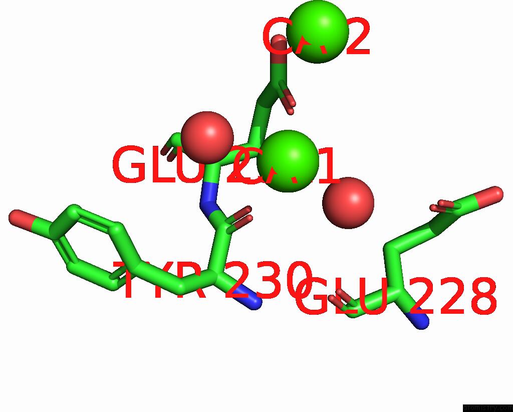
Calcium binding site 1 out of 5 in 3bs6
Go back to
Calcium binding site 1 out
of 5 in the 1.8 Angstrom Crystal Structure of the Periplasmic Domain of the Membrane Insertase Yidc
Mono view

Stereo pair view
Mono view
Stereo pair view
A full contact list of Calcium with other atoms in the Ca binding
site number 1 of 1.8 Angstrom Crystal Structure of the Periplasmic Domain of the Membrane Insertase Yidc within 5.0Å range:
|
Calcium binding site 2 out of 5 in 3bs6
Go back to
Calcium binding site 2 out
of 5 in the 1.8 Angstrom Crystal Structure of the Periplasmic Domain of the Membrane Insertase Yidc

Mono view

Stereo pair view
Mono view
Stereo pair view
A full contact list of Calcium with other atoms in the Ca binding
site number 2 of 1.8 Angstrom Crystal Structure of the Periplasmic Domain of the Membrane Insertase Yidc within 5.0Å range:
|
Calcium binding site 3 out of 5 in 3bs6
Go back to
Calcium binding site 3 out
of 5 in the 1.8 Angstrom Crystal Structure of the Periplasmic Domain of the Membrane Insertase Yidc

Mono view

Stereo pair view
Mono view
Stereo pair view
A full contact list of Calcium with other atoms in the Ca binding
site number 3 of 1.8 Angstrom Crystal Structure of the Periplasmic Domain of the Membrane Insertase Yidc within 5.0Å range:
|
Calcium binding site 4 out of 5 in 3bs6
Go back to
Calcium binding site 4 out
of 5 in the 1.8 Angstrom Crystal Structure of the Periplasmic Domain of the Membrane Insertase Yidc

Mono view

Stereo pair view
Mono view
Stereo pair view
A full contact list of Calcium with other atoms in the Ca binding
site number 4 of 1.8 Angstrom Crystal Structure of the Periplasmic Domain of the Membrane Insertase Yidc within 5.0Å range:
|
Calcium binding site 5 out of 5 in 3bs6
Go back to
Calcium binding site 5 out
of 5 in the 1.8 Angstrom Crystal Structure of the Periplasmic Domain of the Membrane Insertase Yidc

Mono view

Stereo pair view
Mono view
Stereo pair view
A full contact list of Calcium with other atoms in the Ca binding
site number 5 of 1.8 Angstrom Crystal Structure of the Periplasmic Domain of the Membrane Insertase Yidc within 5.0Å range:
|
Reference:
S.Ravaud,
G.Stjepanovic,
K.Wild,
I.Sinning.
The Crystal Structure of the Periplasmic Domain of the Escherichia Coli Membrane Protein Insertase Yidc Contains A Substrate Binding Cleft J.Biol.Chem. V. 283 9350 2008.
ISSN: ISSN 0021-9258
PubMed: 18234665
DOI: 10.1074/JBC.M710493200
Page generated: Tue Jul 8 11:11:15 2025
ISSN: ISSN 0021-9258
PubMed: 18234665
DOI: 10.1074/JBC.M710493200
Last articles
Mg in 8CGIMg in 8CF8
Mg in 8CGA
Mg in 8CEP
Mg in 8CF1
Mg in 8CAH
Mg in 8CE5
Mg in 8CDQ
Mg in 8CE2
Mg in 8CCO