Calcium »
PDB 3bje-3bxk »
3bse »
Calcium in PDB 3bse: Crystal Structure Analysis of A 16-Base-Pair B-Dna
Protein crystallography data
The structure of Crystal Structure Analysis of A 16-Base-Pair B-Dna, PDB code: 3bse
was solved by
N.Narayana,
with X-Ray Crystallography technique. A brief refinement statistics is given in the table below:
| Resolution Low / High (Å) | 8.00 / 1.60 |
| Space group | H 3 |
| Cell size a, b, c (Å), α, β, γ (°) | 38.740, 38.740, 161.327, 90.00, 90.00, 120.00 |
| R / Rfree (%) | 22.1 / 27.4 |
Calcium Binding Sites:
Pages:
>>> Page 1 <<< Page 2, Binding sites: 11 - 12;Binding sites:
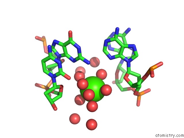
The binding sites of Calcium atom in the Crystal Structure Analysis of A 16-Base-Pair B-Dna (pdb code 3bse). This binding sites where shown within 5.0 Angstroms radius around Calcium atom.In total 12 binding sites of Calcium where determined in the Crystal Structure Analysis of A 16-Base-Pair B-Dna, PDB code: 3bse:
Jump to Calcium binding site number: 1; 2; 3; 4; 5; 6; 7; 8; 9; 10;
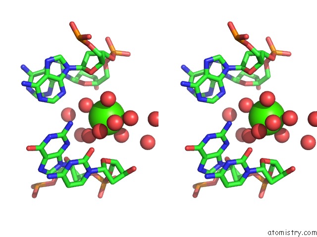
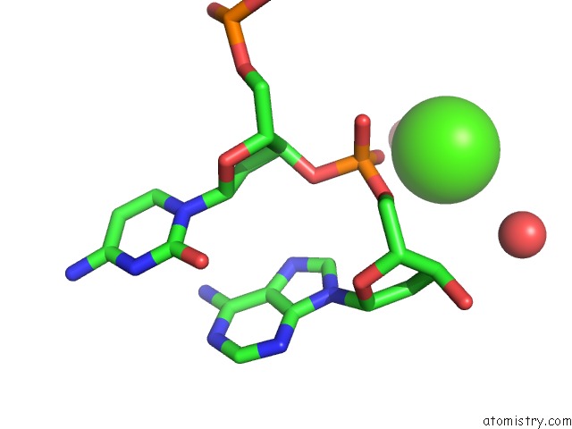
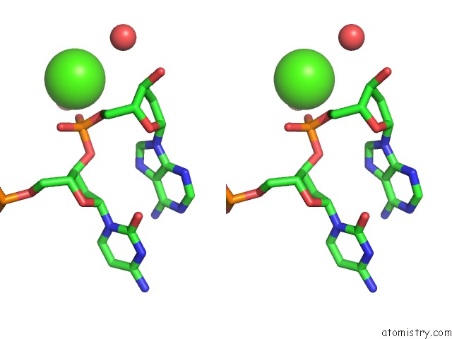
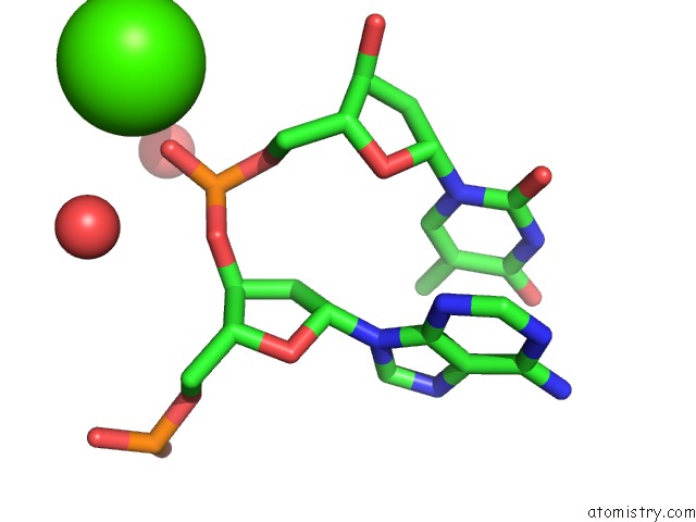
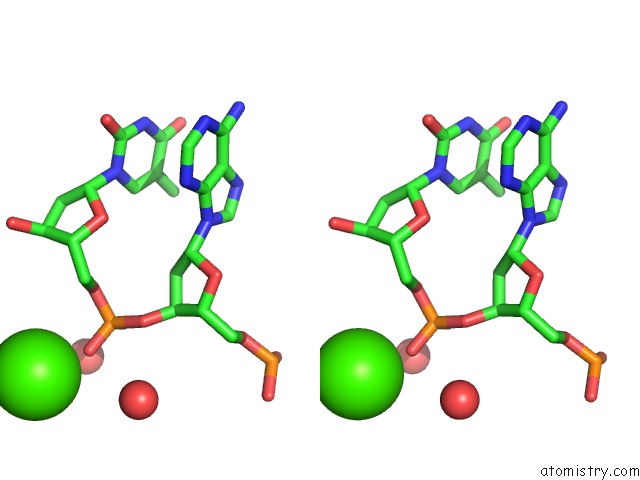
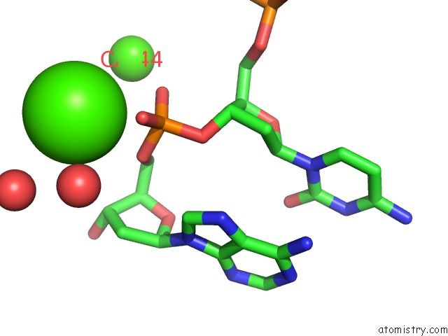
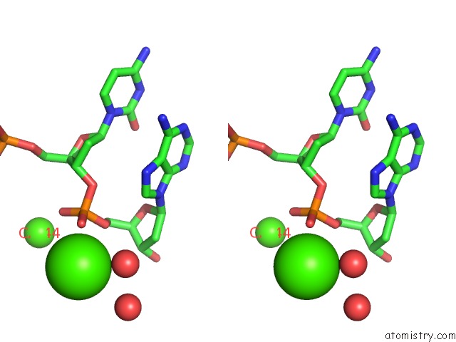
Calcium binding site 1 out of 12 in 3bse
Go back to
Calcium binding site 1 out
of 12 in the Crystal Structure Analysis of A 16-Base-Pair B-Dna
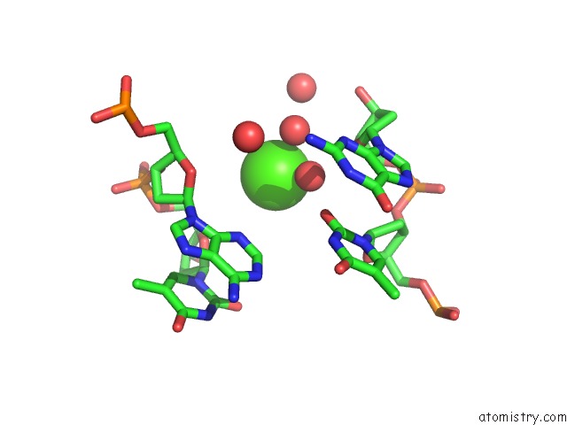
Mono view
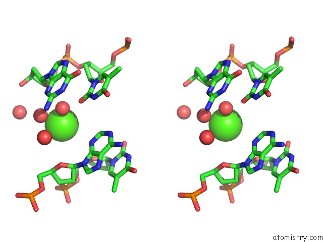
Stereo pair view
Mono view
Stereo pair view
A full contact list of Calcium with other atoms in the Ca binding
site number 1 of Crystal Structure Analysis of A 16-Base-Pair B-Dna within 5.0Å range:
|
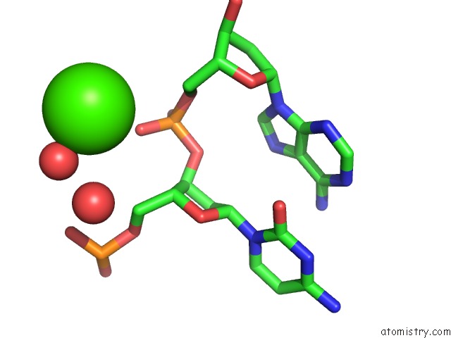
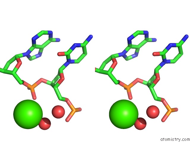
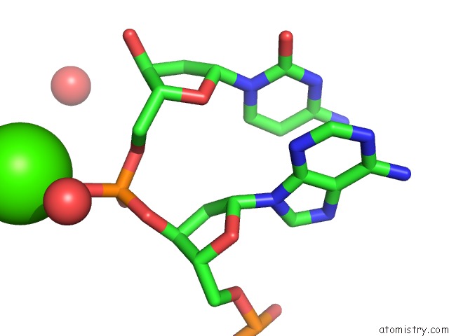
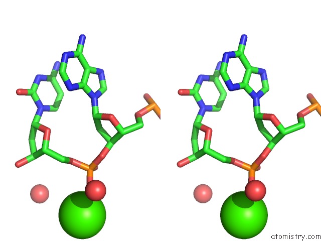
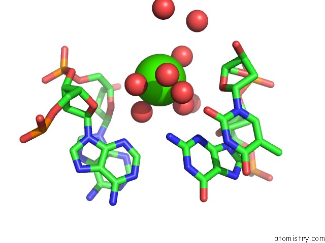
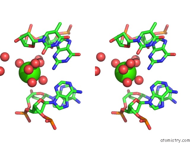
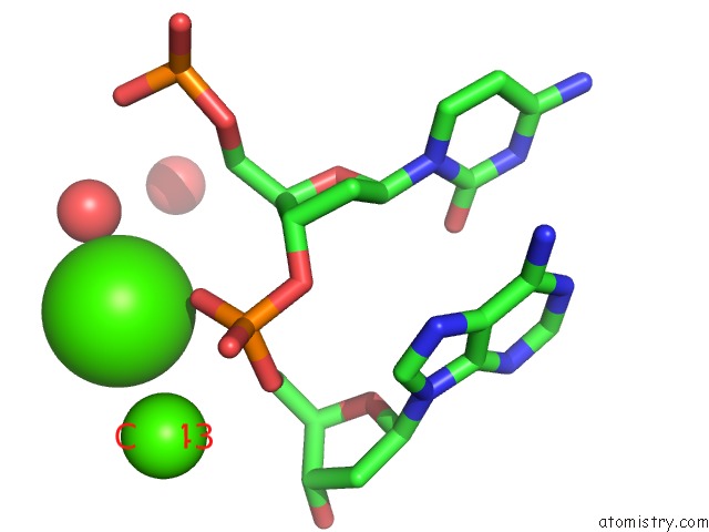
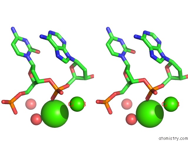
Calcium binding site 2 out of 12 in 3bse
Go back to
Calcium binding site 2 out
of 12 in the Crystal Structure Analysis of A 16-Base-Pair B-Dna
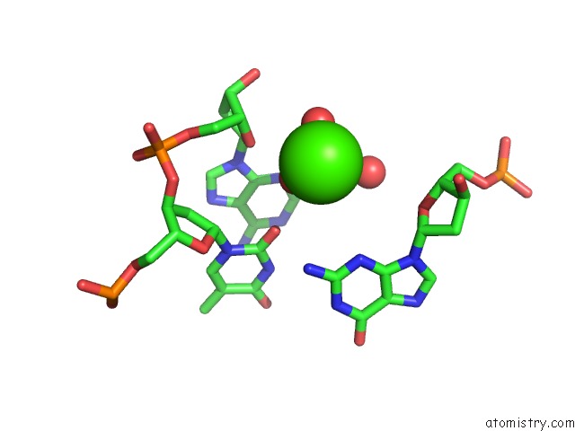
Mono view
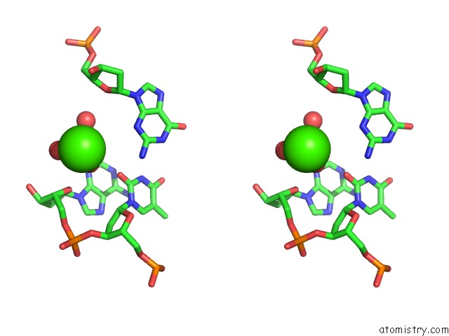
Stereo pair view
Mono view
Stereo pair view
A full contact list of Calcium with other atoms in the Ca binding
site number 2 of Crystal Structure Analysis of A 16-Base-Pair B-Dna within 5.0Å range:
|
Calcium binding site 3 out of 12 in 3bse
Go back to
Calcium binding site 3 out
of 12 in the Crystal Structure Analysis of A 16-Base-Pair B-Dna
Mono view
Stereo pair view
Mono view
Stereo pair view
A full contact list of Calcium with other atoms in the Ca binding
site number 3 of Crystal Structure Analysis of A 16-Base-Pair B-Dna within 5.0Å range:
|
Calcium binding site 4 out of 12 in 3bse
Go back to
Calcium binding site 4 out
of 12 in the Crystal Structure Analysis of A 16-Base-Pair B-Dna
Mono view
Stereo pair view
Mono view
Stereo pair view
A full contact list of Calcium with other atoms in the Ca binding
site number 4 of Crystal Structure Analysis of A 16-Base-Pair B-Dna within 5.0Å range:
|
Calcium binding site 5 out of 12 in 3bse
Go back to
Calcium binding site 5 out
of 12 in the Crystal Structure Analysis of A 16-Base-Pair B-Dna
Mono view
Stereo pair view
Mono view
Stereo pair view
A full contact list of Calcium with other atoms in the Ca binding
site number 5 of Crystal Structure Analysis of A 16-Base-Pair B-Dna within 5.0Å range:
|
Calcium binding site 6 out of 12 in 3bse
Go back to
Calcium binding site 6 out
of 12 in the Crystal Structure Analysis of A 16-Base-Pair B-Dna
Mono view
Stereo pair view
Mono view
Stereo pair view
A full contact list of Calcium with other atoms in the Ca binding
site number 6 of Crystal Structure Analysis of A 16-Base-Pair B-Dna within 5.0Å range:
|
Calcium binding site 7 out of 12 in 3bse
Go back to
Calcium binding site 7 out
of 12 in the Crystal Structure Analysis of A 16-Base-Pair B-Dna
Mono view
Stereo pair view
Mono view
Stereo pair view
A full contact list of Calcium with other atoms in the Ca binding
site number 7 of Crystal Structure Analysis of A 16-Base-Pair B-Dna within 5.0Å range:
|
Calcium binding site 8 out of 12 in 3bse
Go back to
Calcium binding site 8 out
of 12 in the Crystal Structure Analysis of A 16-Base-Pair B-Dna
Mono view
Stereo pair view
Mono view
Stereo pair view
A full contact list of Calcium with other atoms in the Ca binding
site number 8 of Crystal Structure Analysis of A 16-Base-Pair B-Dna within 5.0Å range:
|
Calcium binding site 9 out of 12 in 3bse
Go back to
Calcium binding site 9 out
of 12 in the Crystal Structure Analysis of A 16-Base-Pair B-Dna
Mono view
Stereo pair view
Mono view
Stereo pair view
A full contact list of Calcium with other atoms in the Ca binding
site number 9 of Crystal Structure Analysis of A 16-Base-Pair B-Dna within 5.0Å range:
|
Calcium binding site 10 out of 12 in 3bse
Go back to
Calcium binding site 10 out
of 12 in the Crystal Structure Analysis of A 16-Base-Pair B-Dna
Mono view
Stereo pair view
Mono view
Stereo pair view
A full contact list of Calcium with other atoms in the Ca binding
site number 10 of Crystal Structure Analysis of A 16-Base-Pair B-Dna within 5.0Å range:
|
Reference:
N.Narayana,
M.A.Weiss.
Crystallographic Analysis of A Sex-Specific Enhancer Element: Sequence-Dependent Dna Structure, Hydration, and Dynamics J.Mol.Biol. V. 385 469 2009.
ISSN: ISSN 0022-2836
PubMed: 18992257
DOI: 10.1016/J.JMB.2008.10.041
Page generated: Tue Jul 8 11:11:53 2025
ISSN: ISSN 0022-2836
PubMed: 18992257
DOI: 10.1016/J.JMB.2008.10.041
Last articles
Mg in 8H53Mg in 8H68
Mg in 8H5Y
Mg in 8H4P
Mg in 8H5M
Mg in 8H4G
Mg in 8H4F
Mg in 8H4D
Mg in 8H4E
Mg in 8H4C