Calcium »
PDB 3bxl-3cfw »
3c6l »
Calcium in PDB 3c6l: Crystal Structure of Mouse Mhc Class II I-Ab/3K Peptide Complexed with Mouse Tcr 2W20
Protein crystallography data
The structure of Crystal Structure of Mouse Mhc Class II I-Ab/3K Peptide Complexed with Mouse Tcr 2W20, PDB code: 3c6l
was solved by
S.Dai,
with X-Ray Crystallography technique. A brief refinement statistics is given in the table below:
| Resolution Low / High (Å) | 49.07 / 3.40 |
| Space group | P 21 21 21 |
| Cell size a, b, c (Å), α, β, γ (°) | 46.540, 113.930, 386.260, 90.00, 90.00, 90.00 |
| R / Rfree (%) | 26.8 / 32.5 |
Calcium Binding Sites:
The binding sites of Calcium atom in the Crystal Structure of Mouse Mhc Class II I-Ab/3K Peptide Complexed with Mouse Tcr 2W20
(pdb code 3c6l). This binding sites where shown within
5.0 Angstroms radius around Calcium atom.
In total 3 binding sites of Calcium where determined in the Crystal Structure of Mouse Mhc Class II I-Ab/3K Peptide Complexed with Mouse Tcr 2W20, PDB code: 3c6l:
Jump to Calcium binding site number: 1; 2; 3;
In total 3 binding sites of Calcium where determined in the Crystal Structure of Mouse Mhc Class II I-Ab/3K Peptide Complexed with Mouse Tcr 2W20, PDB code: 3c6l:
Jump to Calcium binding site number: 1; 2; 3;
Calcium binding site 1 out of 3 in 3c6l
Go back to
Calcium binding site 1 out
of 3 in the Crystal Structure of Mouse Mhc Class II I-Ab/3K Peptide Complexed with Mouse Tcr 2W20
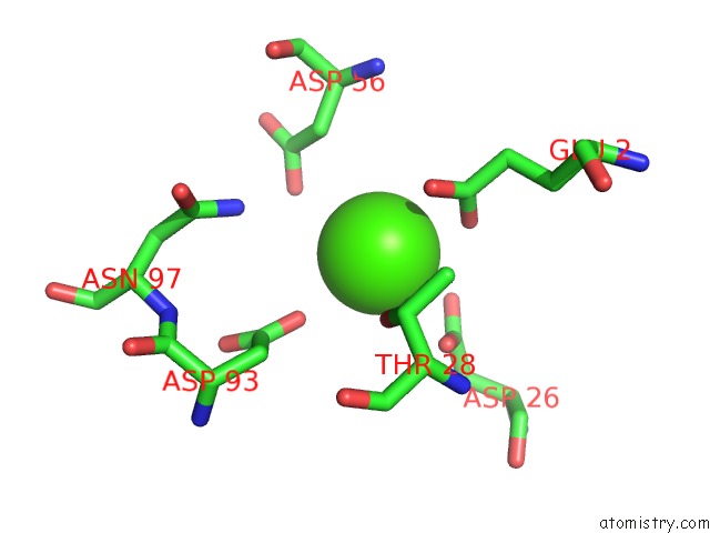
Mono view
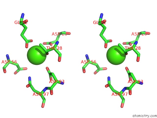
Stereo pair view
Mono view
Stereo pair view
A full contact list of Calcium with other atoms in the Ca binding
site number 1 of Crystal Structure of Mouse Mhc Class II I-Ab/3K Peptide Complexed with Mouse Tcr 2W20 within 5.0Å range:
|
Calcium binding site 2 out of 3 in 3c6l
Go back to
Calcium binding site 2 out
of 3 in the Crystal Structure of Mouse Mhc Class II I-Ab/3K Peptide Complexed with Mouse Tcr 2W20
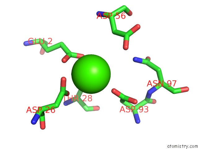
Mono view
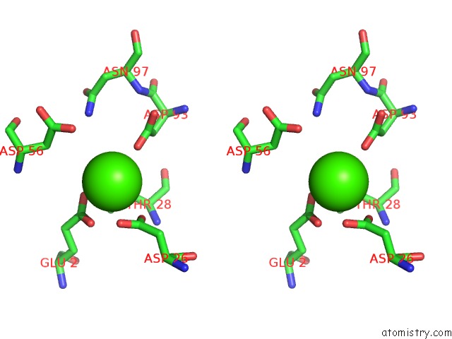
Stereo pair view
Mono view
Stereo pair view
A full contact list of Calcium with other atoms in the Ca binding
site number 2 of Crystal Structure of Mouse Mhc Class II I-Ab/3K Peptide Complexed with Mouse Tcr 2W20 within 5.0Å range:
|
Calcium binding site 3 out of 3 in 3c6l
Go back to
Calcium binding site 3 out
of 3 in the Crystal Structure of Mouse Mhc Class II I-Ab/3K Peptide Complexed with Mouse Tcr 2W20
Mono view
Stereo pair view
Mono view
Stereo pair view
| A full contact list of Calcium with other atoms in the Ca binding site number 3 of Crystal Structure of Mouse Mhc Class II I-Ab/3K Peptide Complexed with Mouse Tcr 2W20 within 5.0Å range: |
Reference:
S.Dai,
E.S.Huseby,
K.Rubtsova,
J.Scott-Browne,
F.Crawford,
W.A.Macdonald,
P.Marrack,
J.W.Kappler.
Crossreactive T Cells Spotlight the Germline Rules For Alphabeta T Cell-Receptor Interactions with Mhc Molecules. Immunity V. 28 324 2008.
ISSN: ISSN 1074-7613
PubMed: 18308592
DOI: 10.1016/J.IMMUNI.2008.01.008
Page generated: Tue Jul 8 11:19:56 2025
ISSN: ISSN 1074-7613
PubMed: 18308592
DOI: 10.1016/J.IMMUNI.2008.01.008
Last articles
I in 4CB6I in 4DCH
I in 4BH5
I in 4BVA
I in 4D85
I in 4CJD
I in 4CDW
I in 4BVX
I in 4BSW
I in 4AW7