Calcium »
PDB 3cu2-3dc0 »
3d5y »
Calcium in PDB 3d5y: High Resolution Crystal Structure of 1,5-Alpha-Arabinanase Catalytic Mutant (ABNBE201A)
Enzymatic activity of High Resolution Crystal Structure of 1,5-Alpha-Arabinanase Catalytic Mutant (ABNBE201A)
All present enzymatic activity of High Resolution Crystal Structure of 1,5-Alpha-Arabinanase Catalytic Mutant (ABNBE201A):
3.2.1.99;
3.2.1.99;
Protein crystallography data
The structure of High Resolution Crystal Structure of 1,5-Alpha-Arabinanase Catalytic Mutant (ABNBE201A), PDB code: 3d5y
was solved by
A.Alhassid,
A.Ben David,
Y.Shoham,
G.Shoham,
with X-Ray Crystallography technique. A brief refinement statistics is given in the table below:
| Resolution Low / High (Å) | 30.00 / 1.22 |
| Space group | C 2 2 21 |
| Cell size a, b, c (Å), α, β, γ (°) | 85.854, 89.672, 75.503, 90.00, 90.00, 90.00 |
| R / Rfree (%) | 15.8 / 19.2 |
Calcium Binding Sites:
The binding sites of Calcium atom in the High Resolution Crystal Structure of 1,5-Alpha-Arabinanase Catalytic Mutant (ABNBE201A)
(pdb code 3d5y). This binding sites where shown within
5.0 Angstroms radius around Calcium atom.
In total only one binding site of Calcium was determined in the High Resolution Crystal Structure of 1,5-Alpha-Arabinanase Catalytic Mutant (ABNBE201A), PDB code: 3d5y:
In total only one binding site of Calcium was determined in the High Resolution Crystal Structure of 1,5-Alpha-Arabinanase Catalytic Mutant (ABNBE201A), PDB code: 3d5y:
Calcium binding site 1 out of 1 in 3d5y
Go back to
Calcium binding site 1 out
of 1 in the High Resolution Crystal Structure of 1,5-Alpha-Arabinanase Catalytic Mutant (ABNBE201A)
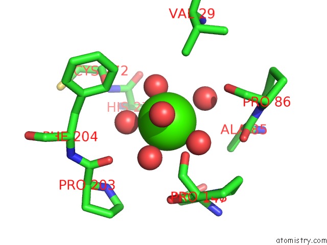
Mono view
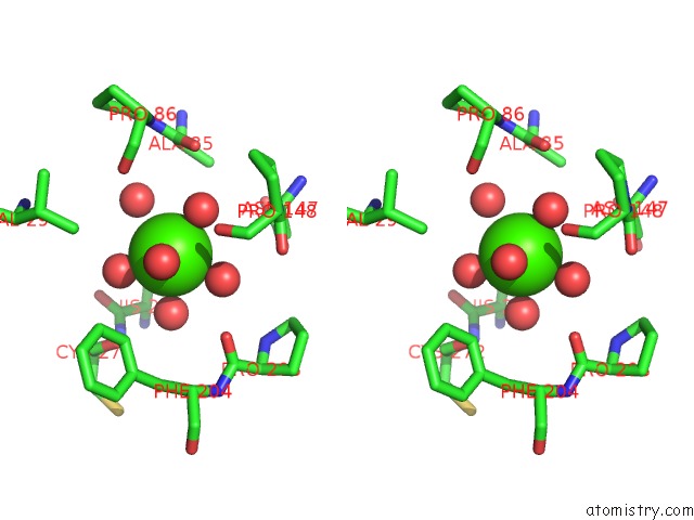
Stereo pair view
Mono view
Stereo pair view
A full contact list of Calcium with other atoms in the Ca binding
site number 1 of High Resolution Crystal Structure of 1,5-Alpha-Arabinanase Catalytic Mutant (ABNBE201A) within 5.0Å range:
|
Reference:
A.Alhassid,
A.Ben-David,
O.Tabachnikov,
D.Libster,
E.Naveh,
G.Zolotnitsky,
Y.Shoham,
G.Shoham.
Crystal Structure of An Inverting Gh 43 1,5-Alpha-L-Arabinanase From Geobacillus Stearothermophilus Complexed with Its Substrate Biochem.J. V. 422 73 2009.
ISSN: ISSN 0264-6021
PubMed: 19505290
DOI: 10.1042/BJ20090180
Page generated: Tue Jul 8 11:31:07 2025
ISSN: ISSN 0264-6021
PubMed: 19505290
DOI: 10.1042/BJ20090180
Last articles
Mg in 4JJSMg in 4JJ2
Mg in 4JIW
Mg in 4JIV
Mg in 4JIB
Mg in 4JI4
Mg in 4JI5
Mg in 4JI1
Mg in 4JI0
Mg in 4JI2