Calcium »
PDB 3cu2-3dc0 »
3d65 »
Calcium in PDB 3d65: Crystal Structure of Textilinin-1, A Kunitz-Type Serine Protease Inhibitor From the Australian Common Brown Snake Venom, in Complex with Trypsin
Enzymatic activity of Crystal Structure of Textilinin-1, A Kunitz-Type Serine Protease Inhibitor From the Australian Common Brown Snake Venom, in Complex with Trypsin
All present enzymatic activity of Crystal Structure of Textilinin-1, A Kunitz-Type Serine Protease Inhibitor From the Australian Common Brown Snake Venom, in Complex with Trypsin:
3.4.21.4;
3.4.21.4;
Protein crystallography data
The structure of Crystal Structure of Textilinin-1, A Kunitz-Type Serine Protease Inhibitor From the Australian Common Brown Snake Venom, in Complex with Trypsin, PDB code: 3d65
was solved by
E.-K.I.Millers,
P.P.Masci,
M.F.Lavin,
J.De Jersey,
L.W.Guddat,
with X-Ray Crystallography technique. A brief refinement statistics is given in the table below:
| Resolution Low / High (Å) | 37.42 / 1.64 |
| Space group | P 31 2 1 |
| Cell size a, b, c (Å), α, β, γ (°) | 79.838, 79.838, 107.394, 90.00, 90.00, 120.00 |
| R / Rfree (%) | 21.3 / 24.5 |
Calcium Binding Sites:
The binding sites of Calcium atom in the Crystal Structure of Textilinin-1, A Kunitz-Type Serine Protease Inhibitor From the Australian Common Brown Snake Venom, in Complex with Trypsin
(pdb code 3d65). This binding sites where shown within
5.0 Angstroms radius around Calcium atom.
In total only one binding site of Calcium was determined in the Crystal Structure of Textilinin-1, A Kunitz-Type Serine Protease Inhibitor From the Australian Common Brown Snake Venom, in Complex with Trypsin, PDB code: 3d65:
In total only one binding site of Calcium was determined in the Crystal Structure of Textilinin-1, A Kunitz-Type Serine Protease Inhibitor From the Australian Common Brown Snake Venom, in Complex with Trypsin, PDB code: 3d65:
Calcium binding site 1 out of 1 in 3d65
Go back to
Calcium binding site 1 out
of 1 in the Crystal Structure of Textilinin-1, A Kunitz-Type Serine Protease Inhibitor From the Australian Common Brown Snake Venom, in Complex with Trypsin
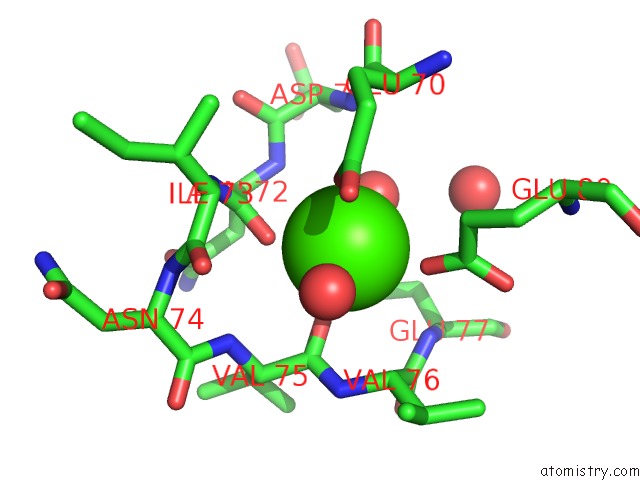
Mono view
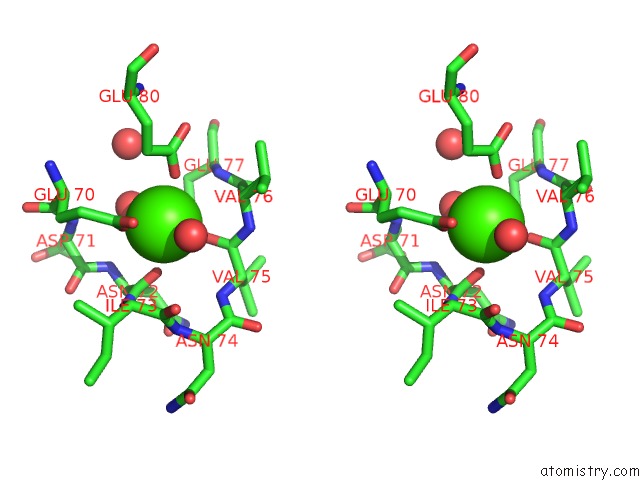
Stereo pair view
Mono view
Stereo pair view
A full contact list of Calcium with other atoms in the Ca binding
site number 1 of Crystal Structure of Textilinin-1, A Kunitz-Type Serine Protease Inhibitor From the Australian Common Brown Snake Venom, in Complex with Trypsin within 5.0Å range:
|
Reference:
E.-K.I.Millers,
M.F.Lavin,
J.De Jersey,
P.P.Masci,
L.W.Guddat.
Crystal Structure of Textilinin-1, A Kunitz-Type Serine Protease Inhibitor From the Australian Common Brown Snake Venom, in Complex with Trypsin To Be Published.
Page generated: Tue Jul 8 11:32:05 2025
Last articles
Mg in 4J9KMg in 4J99
Mg in 4J96
Mg in 4J8W
Mg in 4J8X
Mg in 4J8U
Mg in 4J8V
Mg in 4J8M
Mg in 4J6W
Mg in 4J8F