Calcium »
PDB 3cu2-3dc0 »
3d6c »
Calcium in PDB 3d6c: Crystal Structure of Staphylococcal Nuclease Variant Phs L38E at Cryogenic Temperature
Enzymatic activity of Crystal Structure of Staphylococcal Nuclease Variant Phs L38E at Cryogenic Temperature
All present enzymatic activity of Crystal Structure of Staphylococcal Nuclease Variant Phs L38E at Cryogenic Temperature:
3.1.31.1;
3.1.31.1;
Protein crystallography data
The structure of Crystal Structure of Staphylococcal Nuclease Variant Phs L38E at Cryogenic Temperature, PDB code: 3d6c
was solved by
M.J.Harms,
J.L.Schlessman,
G.R.Sue,
E.B.Garcia-Moreno,
with X-Ray Crystallography technique. A brief refinement statistics is given in the table below:
| Resolution Low / High (Å) | 48.28 / 2.00 |
| Space group | P 41 |
| Cell size a, b, c (Å), α, β, γ (°) | 48.256, 48.256, 62.675, 90.00, 90.00, 90.00 |
| R / Rfree (%) | 17.3 / 23.4 |
Calcium Binding Sites:
The binding sites of Calcium atom in the Crystal Structure of Staphylococcal Nuclease Variant Phs L38E at Cryogenic Temperature
(pdb code 3d6c). This binding sites where shown within
5.0 Angstroms radius around Calcium atom.
In total only one binding site of Calcium was determined in the Crystal Structure of Staphylococcal Nuclease Variant Phs L38E at Cryogenic Temperature, PDB code: 3d6c:
In total only one binding site of Calcium was determined in the Crystal Structure of Staphylococcal Nuclease Variant Phs L38E at Cryogenic Temperature, PDB code: 3d6c:
Calcium binding site 1 out of 1 in 3d6c
Go back to
Calcium binding site 1 out
of 1 in the Crystal Structure of Staphylococcal Nuclease Variant Phs L38E at Cryogenic Temperature
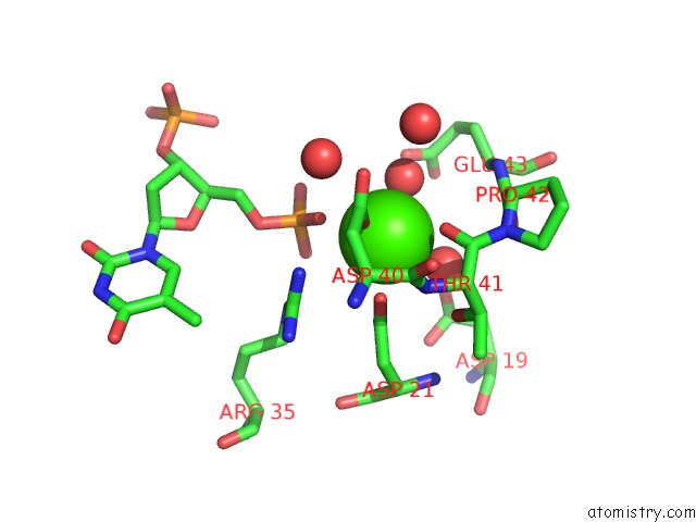
Mono view
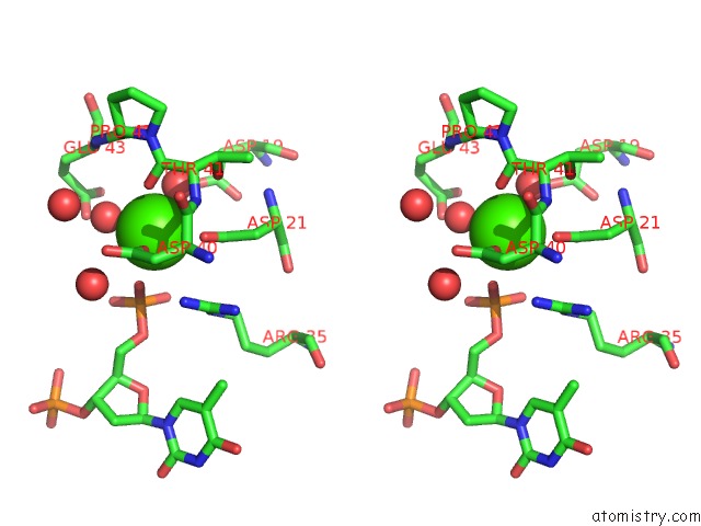
Stereo pair view
Mono view
Stereo pair view
A full contact list of Calcium with other atoms in the Ca binding
site number 1 of Crystal Structure of Staphylococcal Nuclease Variant Phs L38E at Cryogenic Temperature within 5.0Å range:
|
Reference:
M.J.Harms,
C.A.Castaneda,
J.L.Schlessman,
G.R.Sue,
D.G.Isom,
B.R.Cannon,
E.B.Garcia-Moreno.
The Pk(A) Values of Acidic and Basic Residues Buried at the Same Internal Location in A Protein Are Governed By Different Factors. J.Mol.Biol. V. 389 34 2009.
ISSN: ISSN 0022-2836
PubMed: 19324049
DOI: 10.1016/J.JMB.2009.03.039
Page generated: Tue Jul 8 11:32:06 2025
ISSN: ISSN 0022-2836
PubMed: 19324049
DOI: 10.1016/J.JMB.2009.03.039
Last articles
Mg in 4JJSMg in 4JJ2
Mg in 4JIW
Mg in 4JIV
Mg in 4JIB
Mg in 4JI4
Mg in 4JI5
Mg in 4JI1
Mg in 4JI0
Mg in 4JI2