Calcium »
PDB 3eqf-3f45 »
3esq »
Calcium in PDB 3esq: Crystal Structure of Calcium-Bound D,D-Heptose 1.7-Bisphosphate Phosphatase From E. Coli
Protein crystallography data
The structure of Crystal Structure of Calcium-Bound D,D-Heptose 1.7-Bisphosphate Phosphatase From E. Coli, PDB code: 3esq
was solved by
S.N.Sugiman-Marangos,
M.S.Junop,
with X-Ray Crystallography technique. A brief refinement statistics is given in the table below:
| Resolution Low / High (Å) | 40.62 / 1.70 |
| Space group | P 21 21 2 |
| Cell size a, b, c (Å), α, β, γ (°) | 63.904, 50.585, 52.638, 90.00, 90.00, 90.00 |
| R / Rfree (%) | 20 / 25.8 |
Other elements in 3esq:
The structure of Crystal Structure of Calcium-Bound D,D-Heptose 1.7-Bisphosphate Phosphatase From E. Coli also contains other interesting chemical elements:
| Zinc | (Zn) | 1 atom |
Calcium Binding Sites:
The binding sites of Calcium atom in the Crystal Structure of Calcium-Bound D,D-Heptose 1.7-Bisphosphate Phosphatase From E. Coli
(pdb code 3esq). This binding sites where shown within
5.0 Angstroms radius around Calcium atom.
In total only one binding site of Calcium was determined in the Crystal Structure of Calcium-Bound D,D-Heptose 1.7-Bisphosphate Phosphatase From E. Coli, PDB code: 3esq:
In total only one binding site of Calcium was determined in the Crystal Structure of Calcium-Bound D,D-Heptose 1.7-Bisphosphate Phosphatase From E. Coli, PDB code: 3esq:
Calcium binding site 1 out of 1 in 3esq
Go back to
Calcium binding site 1 out
of 1 in the Crystal Structure of Calcium-Bound D,D-Heptose 1.7-Bisphosphate Phosphatase From E. Coli
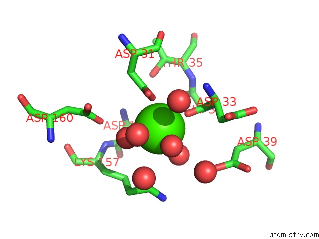
Mono view
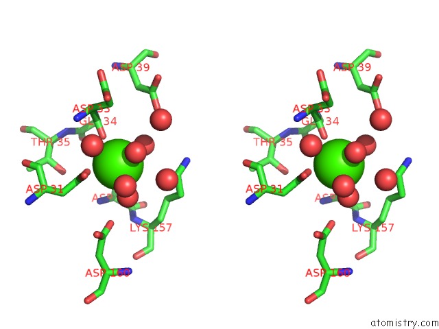
Stereo pair view
Mono view
Stereo pair view
A full contact list of Calcium with other atoms in the Ca binding
site number 1 of Crystal Structure of Calcium-Bound D,D-Heptose 1.7-Bisphosphate Phosphatase From E. Coli within 5.0Å range:
|
Reference:
P.Taylor,
S.N.Sugiman-Marangos,
K.Zhang,
G.Deleon,
G.D.Wright,
M.S.Junop.
Crystal Structure of D,D-Heptose 1.7-Bisphosphate Phosphatase From E. Coli. To Be Published.
Page generated: Tue Jul 8 11:59:57 2025
Last articles
Mg in 6FDIMg in 6FDW
Mg in 6FDS
Mg in 6FD4
Mg in 6FD5
Mg in 6FD6
Mg in 6FD3
Mg in 6FCT
Mg in 6FCW
Mg in 6FCY