Calcium »
PDB 3eqf-3f45 »
3ewt »
Calcium in PDB 3ewt: Crystal Structure of Calmodulin Complexed with A Peptide
Protein crystallography data
The structure of Crystal Structure of Calmodulin Complexed with A Peptide, PDB code: 3ewt
was solved by
T.Jiang,
P.Cao,
Y.Gong,
H.J.Yu,
W.J.Gui,
W.T.Zhang,
with X-Ray Crystallography technique. A brief refinement statistics is given in the table below:
| Resolution Low / High (Å) | 19.90 / 2.40 |
| Space group | P 32 2 1 |
| Cell size a, b, c (Å), α, β, γ (°) | 40.062, 40.062, 174.431, 90.00, 90.00, 120.00 |
| R / Rfree (%) | 21.7 / 25.9 |
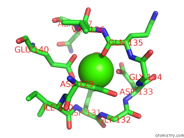
Calcium Binding Sites:
The binding sites of Calcium atom in the Crystal Structure of Calmodulin Complexed with A Peptide
(pdb code 3ewt). This binding sites where shown within
5.0 Angstroms radius around Calcium atom.
In total 4 binding sites of Calcium where determined in the Crystal Structure of Calmodulin Complexed with A Peptide, PDB code: 3ewt:
Jump to Calcium binding site number: 1; 2; 3; 4;
In total 4 binding sites of Calcium where determined in the Crystal Structure of Calmodulin Complexed with A Peptide, PDB code: 3ewt:
Jump to Calcium binding site number: 1; 2; 3; 4;
Calcium binding site 1 out of 4 in 3ewt
Go back to
Calcium binding site 1 out
of 4 in the Crystal Structure of Calmodulin Complexed with A Peptide
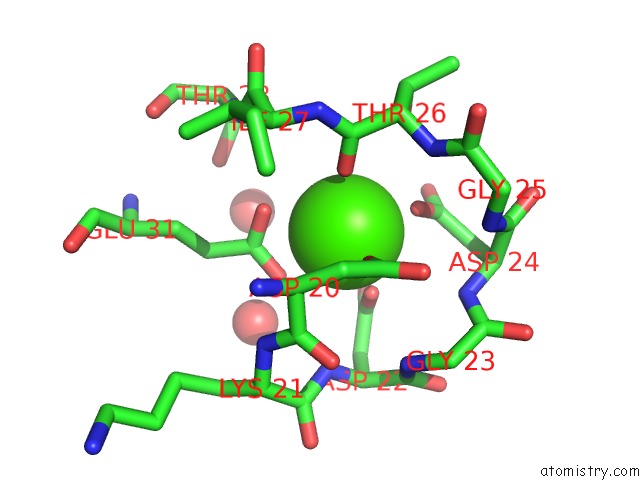
Mono view
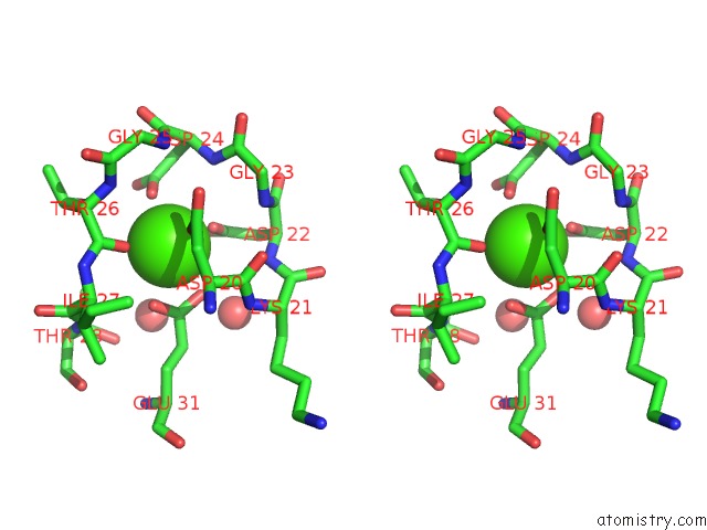
Stereo pair view
Mono view
Stereo pair view
A full contact list of Calcium with other atoms in the Ca binding
site number 1 of Crystal Structure of Calmodulin Complexed with A Peptide within 5.0Å range:
|
Calcium binding site 2 out of 4 in 3ewt
Go back to
Calcium binding site 2 out
of 4 in the Crystal Structure of Calmodulin Complexed with A Peptide
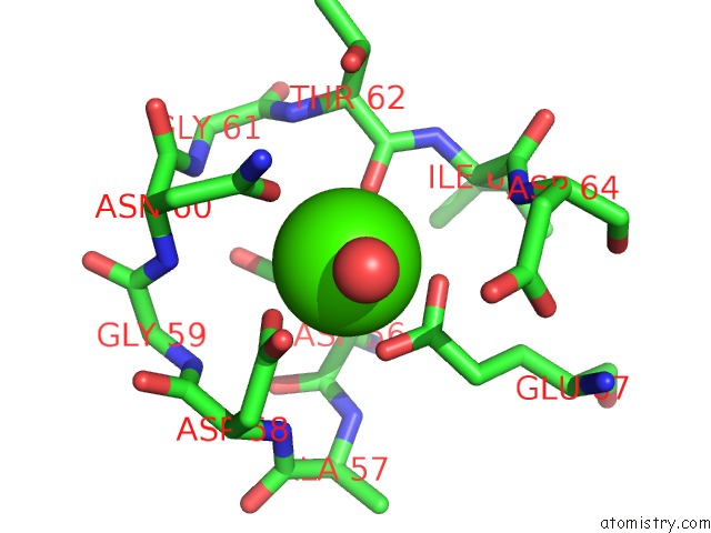
Mono view

Stereo pair view
Mono view
Stereo pair view
A full contact list of Calcium with other atoms in the Ca binding
site number 2 of Crystal Structure of Calmodulin Complexed with A Peptide within 5.0Å range:
|
Calcium binding site 3 out of 4 in 3ewt
Go back to
Calcium binding site 3 out
of 4 in the Crystal Structure of Calmodulin Complexed with A Peptide

Mono view
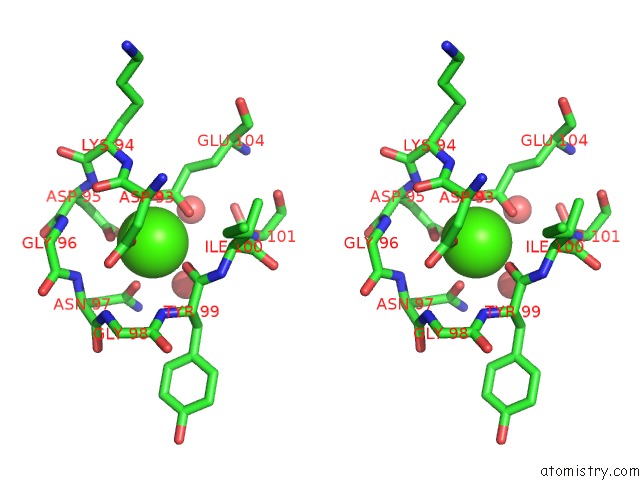
Stereo pair view
Mono view
Stereo pair view
A full contact list of Calcium with other atoms in the Ca binding
site number 3 of Crystal Structure of Calmodulin Complexed with A Peptide within 5.0Å range:
|
Calcium binding site 4 out of 4 in 3ewt
Go back to
Calcium binding site 4 out
of 4 in the Crystal Structure of Calmodulin Complexed with A Peptide
Mono view

Stereo pair view
Mono view
Stereo pair view
A full contact list of Calcium with other atoms in the Ca binding
site number 4 of Crystal Structure of Calmodulin Complexed with A Peptide within 5.0Å range:
|
Reference:
P.Cao,
W.Zhang,
W.Gui,
Y.Dong,
T.Jiang,
Y.Gong.
Structural Insights Into the Mechanism of Calmodulin Binding to Death Receptors. Acta Crystallogr.,Sect.D V. 70 1604 2014.
ISSN: ESSN 1399-0047
PubMed: 24914971
DOI: 10.1107/S1399004714006919
Page generated: Tue Jul 8 12:02:15 2025
ISSN: ESSN 1399-0047
PubMed: 24914971
DOI: 10.1107/S1399004714006919
Last articles
Mg in 6KI8Mg in 6KJ6
Mg in 6KF9
Mg in 6KE2
Mg in 6KF4
Mg in 6KF3
Mg in 6KE4
Mg in 6KE0
Mg in 6KDZ
Mg in 6KDX