Calcium »
PDB 3fyi-3ghg »
3fz5 »
Calcium in PDB 3fz5: Crystal Structure of Possible 2-Hydroxychromene-2-Carboxylate Isomerase From Rhodobacter Sphaeroides
Protein crystallography data
The structure of Crystal Structure of Possible 2-Hydroxychromene-2-Carboxylate Isomerase From Rhodobacter Sphaeroides, PDB code: 3fz5
was solved by
C.Chang,
C.Hatzos,
L.Freeman,
A.Joachimiak,
Midwest Center For Structuralgenomics (Mcsg),
with X-Ray Crystallography technique. A brief refinement statistics is given in the table below:
| Resolution Low / High (Å) | 50.00 / 2.40 |
| Space group | P 1 |
| Cell size a, b, c (Å), α, β, γ (°) | 47.769, 48.724, 95.206, 94.61, 100.46, 90.10 |
| R / Rfree (%) | 20.4 / 26 |
Calcium Binding Sites:
The binding sites of Calcium atom in the Crystal Structure of Possible 2-Hydroxychromene-2-Carboxylate Isomerase From Rhodobacter Sphaeroides
(pdb code 3fz5). This binding sites where shown within
5.0 Angstroms radius around Calcium atom.
In total 2 binding sites of Calcium where determined in the Crystal Structure of Possible 2-Hydroxychromene-2-Carboxylate Isomerase From Rhodobacter Sphaeroides, PDB code: 3fz5:
Jump to Calcium binding site number: 1; 2;
In total 2 binding sites of Calcium where determined in the Crystal Structure of Possible 2-Hydroxychromene-2-Carboxylate Isomerase From Rhodobacter Sphaeroides, PDB code: 3fz5:
Jump to Calcium binding site number: 1; 2;
Calcium binding site 1 out of 2 in 3fz5
Go back to
Calcium binding site 1 out
of 2 in the Crystal Structure of Possible 2-Hydroxychromene-2-Carboxylate Isomerase From Rhodobacter Sphaeroides
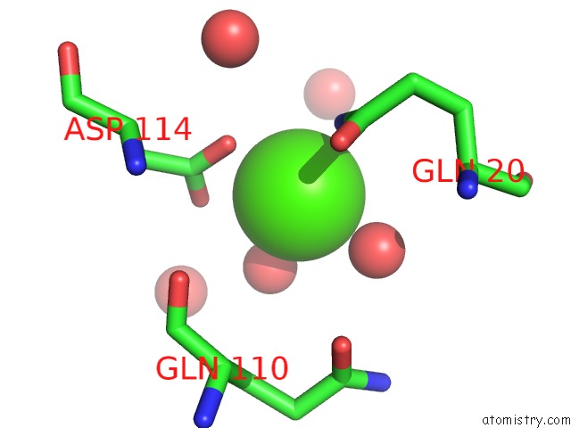
Mono view
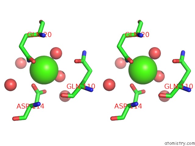
Stereo pair view
Mono view
Stereo pair view
A full contact list of Calcium with other atoms in the Ca binding
site number 1 of Crystal Structure of Possible 2-Hydroxychromene-2-Carboxylate Isomerase From Rhodobacter Sphaeroides within 5.0Å range:
|
Calcium binding site 2 out of 2 in 3fz5
Go back to
Calcium binding site 2 out
of 2 in the Crystal Structure of Possible 2-Hydroxychromene-2-Carboxylate Isomerase From Rhodobacter Sphaeroides
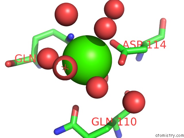
Mono view
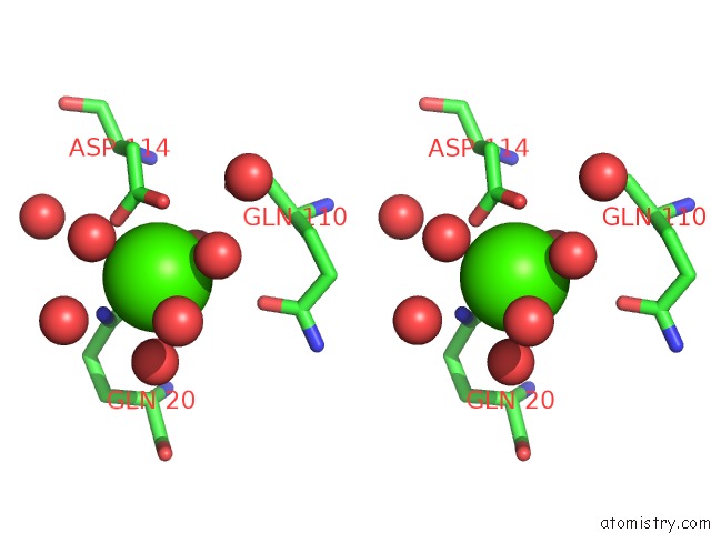
Stereo pair view
Mono view
Stereo pair view
A full contact list of Calcium with other atoms in the Ca binding
site number 2 of Crystal Structure of Possible 2-Hydroxychromene-2-Carboxylate Isomerase From Rhodobacter Sphaeroides within 5.0Å range:
|
Reference:
C.Chang,
C.Hatzos,
L.Freeman,
A.Joachimiak.
Crystal Structure of Possible 2-Hydroxychromene-2-Carboxylate Isomerase From Rhodobacter Sphaeroides To Be Published.
Page generated: Tue Jul 8 12:34:49 2025
Last articles
Na in 8GT0Na in 8H2B
Na in 8GZ1
Na in 8H1G
Na in 8H1B
Na in 8GYR
Na in 8GVM
Na in 8GR6
Na in 8GQ0
Na in 8GSG