Calcium »
PDB 3gif-3gy5 »
3gis »
Calcium in PDB 3gis: Crystal Structure of Na-Free Thrombin in Complex with Thrombomodulin
Enzymatic activity of Crystal Structure of Na-Free Thrombin in Complex with Thrombomodulin
All present enzymatic activity of Crystal Structure of Na-Free Thrombin in Complex with Thrombomodulin:
3.4.21.5;
3.4.21.5;
Protein crystallography data
The structure of Crystal Structure of Na-Free Thrombin in Complex with Thrombomodulin, PDB code: 3gis
was solved by
T.E.Adams,
J.A.Huntington,
with X-Ray Crystallography technique. A brief refinement statistics is given in the table below:
| Resolution Low / High (Å) | 40.00 / 2.40 |
| Space group | P 21 21 21 |
| Cell size a, b, c (Å), α, β, γ (°) | 66.250, 100.340, 229.280, 90.00, 90.00, 90.00 |
| R / Rfree (%) | 21.1 / 25.9 |
Calcium Binding Sites:
The binding sites of Calcium atom in the Crystal Structure of Na-Free Thrombin in Complex with Thrombomodulin
(pdb code 3gis). This binding sites where shown within
5.0 Angstroms radius around Calcium atom.
In total 3 binding sites of Calcium where determined in the Crystal Structure of Na-Free Thrombin in Complex with Thrombomodulin, PDB code: 3gis:
Jump to Calcium binding site number: 1; 2; 3;
In total 3 binding sites of Calcium where determined in the Crystal Structure of Na-Free Thrombin in Complex with Thrombomodulin, PDB code: 3gis:
Jump to Calcium binding site number: 1; 2; 3;
Calcium binding site 1 out of 3 in 3gis
Go back to
Calcium binding site 1 out
of 3 in the Crystal Structure of Na-Free Thrombin in Complex with Thrombomodulin
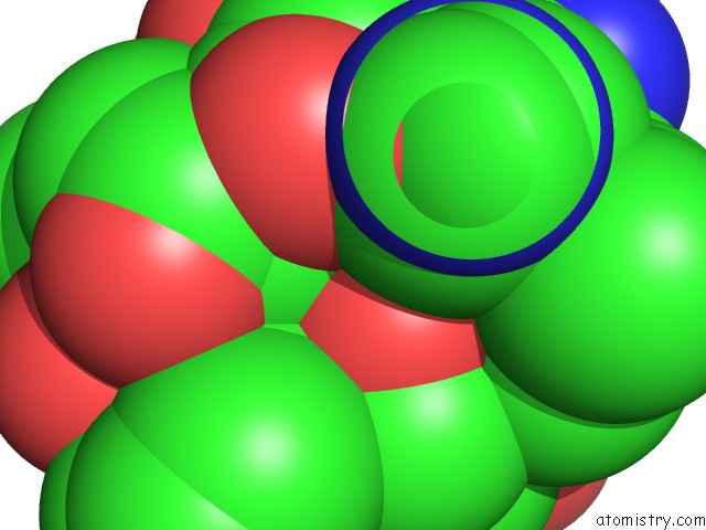
Mono view
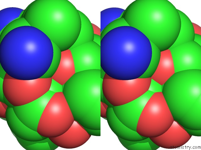
Stereo pair view
Mono view
Stereo pair view
A full contact list of Calcium with other atoms in the Ca binding
site number 1 of Crystal Structure of Na-Free Thrombin in Complex with Thrombomodulin within 5.0Å range:
|
Calcium binding site 2 out of 3 in 3gis
Go back to
Calcium binding site 2 out
of 3 in the Crystal Structure of Na-Free Thrombin in Complex with Thrombomodulin
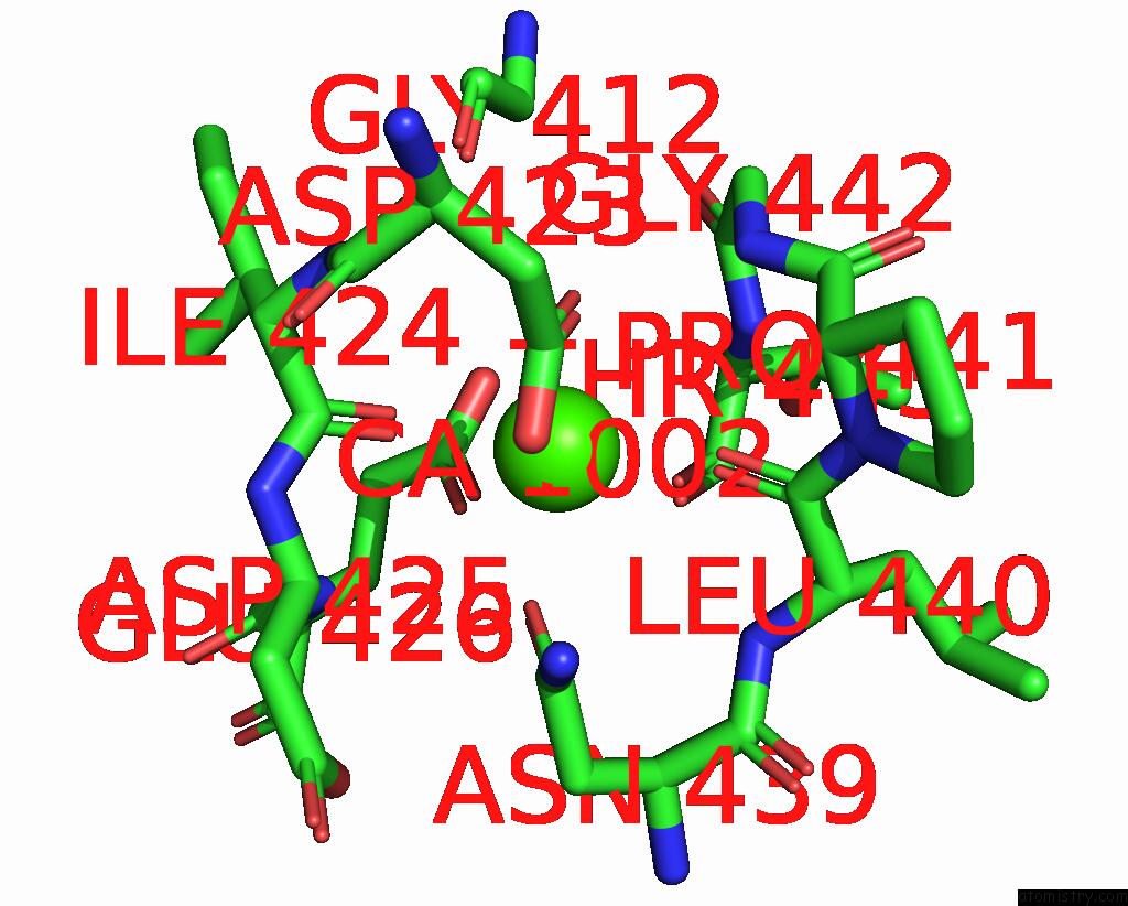
Mono view

Stereo pair view
Mono view
Stereo pair view
A full contact list of Calcium with other atoms in the Ca binding
site number 2 of Crystal Structure of Na-Free Thrombin in Complex with Thrombomodulin within 5.0Å range:
|
Calcium binding site 3 out of 3 in 3gis
Go back to
Calcium binding site 3 out
of 3 in the Crystal Structure of Na-Free Thrombin in Complex with Thrombomodulin
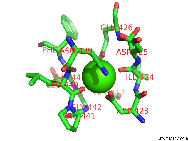
Mono view

Stereo pair view
Mono view
Stereo pair view
A full contact list of Calcium with other atoms in the Ca binding
site number 3 of Crystal Structure of Na-Free Thrombin in Complex with Thrombomodulin within 5.0Å range:
|
Reference:
T.E.Adams,
W.Li,
J.A.Huntington.
Molecular Basis of Thrombomodulin Activation of Slow Thrombin J.Thromb.Haemost. V. 7 1688 2009.
ISSN: ISSN 1538-7933
PubMed: 19656282
DOI: 10.1111/J.1538-7836.2009.03563.X
Page generated: Tue Jul 8 12:45:09 2025
ISSN: ISSN 1538-7933
PubMed: 19656282
DOI: 10.1111/J.1538-7836.2009.03563.X
Last articles
Mg in 5MDLMg in 5MDK
Mg in 5MAC
Mg in 5MDJ
Mg in 5MBK
Mg in 5MAQ
Mg in 5MB9
Mg in 5M8G
Mg in 5M8D
Mg in 5M8B