Calcium »
PDB 3gif-3gy5 »
3gqf »
Calcium in PDB 3gqf: Structural and Biophysical Properties of the Pathogenic SOD1 Variant H46R/H48Q
Enzymatic activity of Structural and Biophysical Properties of the Pathogenic SOD1 Variant H46R/H48Q
All present enzymatic activity of Structural and Biophysical Properties of the Pathogenic SOD1 Variant H46R/H48Q:
1.15.1.1;
1.15.1.1;
Protein crystallography data
The structure of Structural and Biophysical Properties of the Pathogenic SOD1 Variant H46R/H48Q, PDB code: 3gqf
was solved by
D.D.Winkler,
J.P.Schuermann,
P.J.Hart,
with X-Ray Crystallography technique. A brief refinement statistics is given in the table below:
| Resolution Low / High (Å) | 28.07 / 2.20 |
| Space group | C 2 2 21 |
| Cell size a, b, c (Å), α, β, γ (°) | 112.576, 194.331, 143.179, 90.00, 90.00, 90.00 |
| R / Rfree (%) | 20.5 / 24.2 |
Other elements in 3gqf:
The structure of Structural and Biophysical Properties of the Pathogenic SOD1 Variant H46R/H48Q also contains other interesting chemical elements:
| Zinc | (Zn) | 6 atoms |
Calcium Binding Sites:
The binding sites of Calcium atom in the Structural and Biophysical Properties of the Pathogenic SOD1 Variant H46R/H48Q
(pdb code 3gqf). This binding sites where shown within
5.0 Angstroms radius around Calcium atom.
In total 5 binding sites of Calcium where determined in the Structural and Biophysical Properties of the Pathogenic SOD1 Variant H46R/H48Q, PDB code: 3gqf:
Jump to Calcium binding site number: 1; 2; 3; 4; 5;
In total 5 binding sites of Calcium where determined in the Structural and Biophysical Properties of the Pathogenic SOD1 Variant H46R/H48Q, PDB code: 3gqf:
Jump to Calcium binding site number: 1; 2; 3; 4; 5;
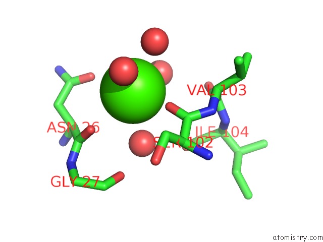
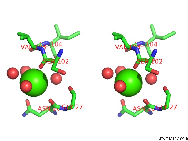
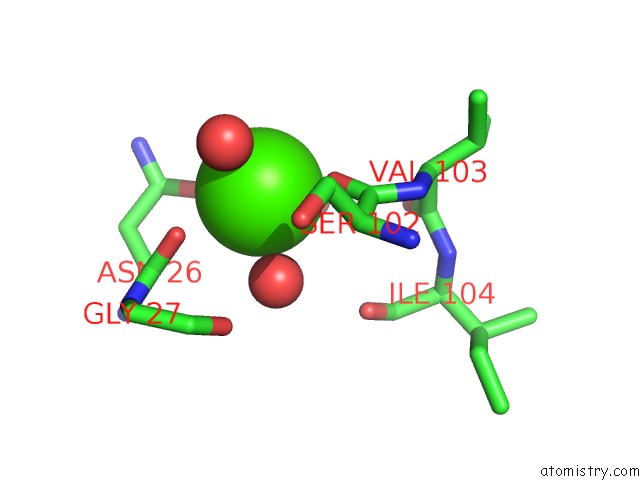
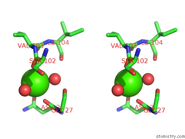
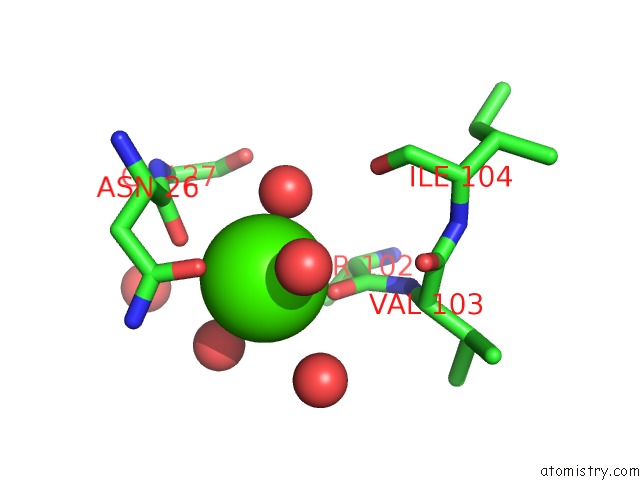
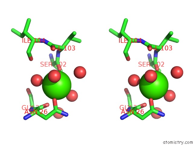
Calcium binding site 1 out of 5 in 3gqf
Go back to
Calcium binding site 1 out
of 5 in the Structural and Biophysical Properties of the Pathogenic SOD1 Variant H46R/H48Q
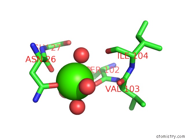
Mono view
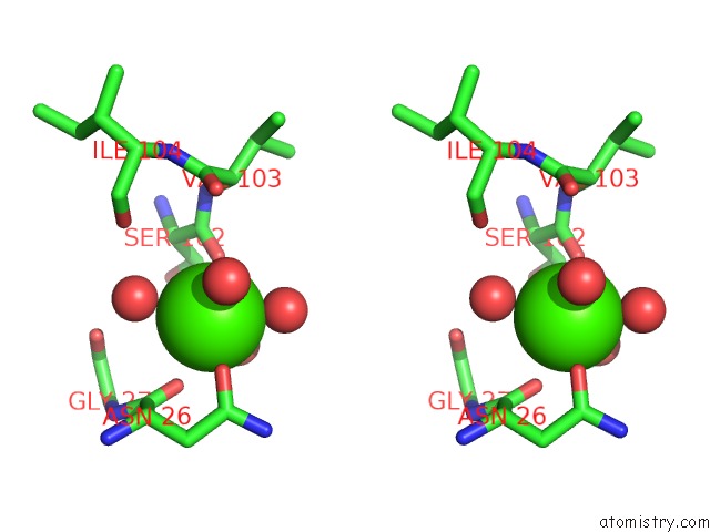
Stereo pair view
Mono view
Stereo pair view
A full contact list of Calcium with other atoms in the Ca binding
site number 1 of Structural and Biophysical Properties of the Pathogenic SOD1 Variant H46R/H48Q within 5.0Å range:
|
Calcium binding site 2 out of 5 in 3gqf
Go back to
Calcium binding site 2 out
of 5 in the Structural and Biophysical Properties of the Pathogenic SOD1 Variant H46R/H48Q
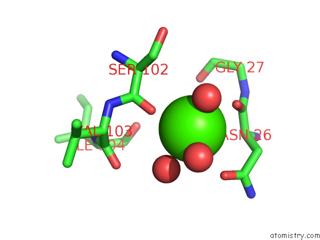
Mono view
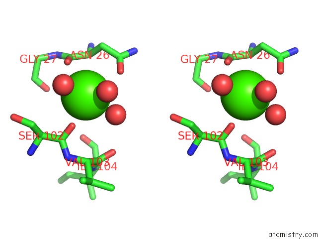
Stereo pair view
Mono view
Stereo pair view
A full contact list of Calcium with other atoms in the Ca binding
site number 2 of Structural and Biophysical Properties of the Pathogenic SOD1 Variant H46R/H48Q within 5.0Å range:
|
Calcium binding site 3 out of 5 in 3gqf
Go back to
Calcium binding site 3 out
of 5 in the Structural and Biophysical Properties of the Pathogenic SOD1 Variant H46R/H48Q
Mono view
Stereo pair view
Mono view
Stereo pair view
A full contact list of Calcium with other atoms in the Ca binding
site number 3 of Structural and Biophysical Properties of the Pathogenic SOD1 Variant H46R/H48Q within 5.0Å range:
|
Calcium binding site 4 out of 5 in 3gqf
Go back to
Calcium binding site 4 out
of 5 in the Structural and Biophysical Properties of the Pathogenic SOD1 Variant H46R/H48Q
Mono view
Stereo pair view
Mono view
Stereo pair view
A full contact list of Calcium with other atoms in the Ca binding
site number 4 of Structural and Biophysical Properties of the Pathogenic SOD1 Variant H46R/H48Q within 5.0Å range:
|
Calcium binding site 5 out of 5 in 3gqf
Go back to
Calcium binding site 5 out
of 5 in the Structural and Biophysical Properties of the Pathogenic SOD1 Variant H46R/H48Q
Mono view
Stereo pair view
Mono view
Stereo pair view
A full contact list of Calcium with other atoms in the Ca binding
site number 5 of Structural and Biophysical Properties of the Pathogenic SOD1 Variant H46R/H48Q within 5.0Å range:
|
Reference:
D.D.Winkler,
J.P.Schuermann,
X.Cao,
S.P.Holloway,
D.R.Borchelt,
M.C.Carroll,
J.B.Proescher,
V.C.Culotta,
P.J.Hart.
Structural and Biophysical Properties of the Pathogenic SOD1 Variant H46R/H48Q. Biochemistry V. 48 3436 2009.
ISSN: ISSN 0006-2960
PubMed: 19227972
DOI: 10.1021/BI8021735
Page generated: Tue Jul 8 12:48:45 2025
ISSN: ISSN 0006-2960
PubMed: 19227972
DOI: 10.1021/BI8021735
Last articles
Mg in 5MH5Mg in 5MGY
Mg in 5MFM
Mg in 5MH1
Mg in 5MGA
Mg in 5M73
Mg in 5MFL
Mg in 5MF4
Mg in 5MF5
Mg in 5MCP