Calcium »
PDB 3gy6-3hii »
3h6g »
Calcium in PDB 3h6g: Crystal Structure of the GLUR6 Amino Terminal Domain Dimer Assembly
Protein crystallography data
The structure of Crystal Structure of the GLUR6 Amino Terminal Domain Dimer Assembly, PDB code: 3h6g
was solved by
J.Kumar,
M.L.Mayer,
with X-Ray Crystallography technique. A brief refinement statistics is given in the table below:
| Resolution Low / High (Å) | 46.80 / 2.70 |
| Space group | P 61 |
| Cell size a, b, c (Å), α, β, γ (°) | 172.074, 172.074, 111.547, 90.00, 90.00, 120.00 |
| R / Rfree (%) | 20.4 / 22.7 |
Calcium Binding Sites:
The binding sites of Calcium atom in the Crystal Structure of the GLUR6 Amino Terminal Domain Dimer Assembly
(pdb code 3h6g). This binding sites where shown within
5.0 Angstroms radius around Calcium atom.
In total 2 binding sites of Calcium where determined in the Crystal Structure of the GLUR6 Amino Terminal Domain Dimer Assembly, PDB code: 3h6g:
Jump to Calcium binding site number: 1; 2;
In total 2 binding sites of Calcium where determined in the Crystal Structure of the GLUR6 Amino Terminal Domain Dimer Assembly, PDB code: 3h6g:
Jump to Calcium binding site number: 1; 2;
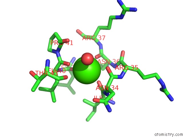
Calcium binding site 1 out of 2 in 3h6g
Go back to
Calcium binding site 1 out
of 2 in the Crystal Structure of the GLUR6 Amino Terminal Domain Dimer Assembly

Mono view

Stereo pair view
Mono view
Stereo pair view
A full contact list of Calcium with other atoms in the Ca binding
site number 1 of Crystal Structure of the GLUR6 Amino Terminal Domain Dimer Assembly within 5.0Å range:
|
Calcium binding site 2 out of 2 in 3h6g
Go back to
Calcium binding site 2 out
of 2 in the Crystal Structure of the GLUR6 Amino Terminal Domain Dimer Assembly
Mono view

Stereo pair view
Mono view
Stereo pair view
A full contact list of Calcium with other atoms in the Ca binding
site number 2 of Crystal Structure of the GLUR6 Amino Terminal Domain Dimer Assembly within 5.0Å range:
|
Reference:
J.Kumar,
P.Schuck,
R.Jin,
M.L.Mayer.
The N-Terminal Domain of GLUR6-Subtype Glutamate Receptor Ion Channels. Nat.Struct.Mol.Biol. V. 16 631 2009.
ISSN: ISSN 1545-9993
PubMed: 19465914
DOI: 10.1038/NSMB.1613
Page generated: Tue Jul 8 12:52:08 2025
ISSN: ISSN 1545-9993
PubMed: 19465914
DOI: 10.1038/NSMB.1613
Last articles
Mg in 6KI8Mg in 6KJ6
Mg in 6KF9
Mg in 6KE2
Mg in 6KF4
Mg in 6KF3
Mg in 6KE4
Mg in 6KE0
Mg in 6KDZ
Mg in 6KDX