Calcium »
PDB 3gy6-3hii »
3h7d »
Calcium in PDB 3h7d: The Crystal Structure of the Cathepsin K Variant M5 in Complex with Chondroitin-4-Sulfate
Enzymatic activity of The Crystal Structure of the Cathepsin K Variant M5 in Complex with Chondroitin-4-Sulfate
All present enzymatic activity of The Crystal Structure of the Cathepsin K Variant M5 in Complex with Chondroitin-4-Sulfate:
3.4.22.38;
3.4.22.38;
Protein crystallography data
The structure of The Crystal Structure of the Cathepsin K Variant M5 in Complex with Chondroitin-4-Sulfate, PDB code: 3h7d
was solved by
M.M.Cherney,
M.Kienetz,
D.Bromme,
M.N.G.James,
with X-Ray Crystallography technique. A brief refinement statistics is given in the table below:
| Resolution Low / High (Å) | 40.25 / 2.24 |
| Space group | C 1 2 1 |
| Cell size a, b, c (Å), α, β, γ (°) | 140.564, 42.014, 87.509, 90.00, 94.19, 90.00 |
| R / Rfree (%) | 17.7 / 23.8 |
Calcium Binding Sites:
The binding sites of Calcium atom in the The Crystal Structure of the Cathepsin K Variant M5 in Complex with Chondroitin-4-Sulfate
(pdb code 3h7d). This binding sites where shown within
5.0 Angstroms radius around Calcium atom.
In total 2 binding sites of Calcium where determined in the The Crystal Structure of the Cathepsin K Variant M5 in Complex with Chondroitin-4-Sulfate, PDB code: 3h7d:
Jump to Calcium binding site number: 1; 2;
In total 2 binding sites of Calcium where determined in the The Crystal Structure of the Cathepsin K Variant M5 in Complex with Chondroitin-4-Sulfate, PDB code: 3h7d:
Jump to Calcium binding site number: 1; 2;
Calcium binding site 1 out of 2 in 3h7d
Go back to
Calcium binding site 1 out
of 2 in the The Crystal Structure of the Cathepsin K Variant M5 in Complex with Chondroitin-4-Sulfate
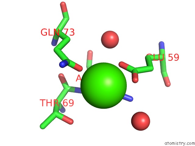
Mono view
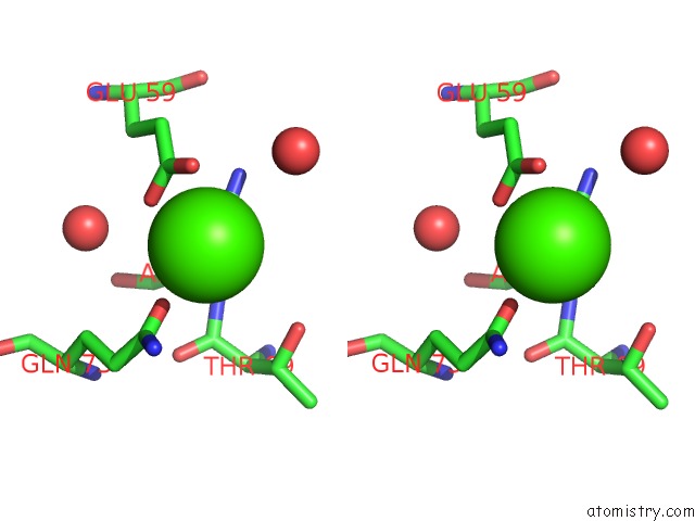
Stereo pair view
Mono view
Stereo pair view
A full contact list of Calcium with other atoms in the Ca binding
site number 1 of The Crystal Structure of the Cathepsin K Variant M5 in Complex with Chondroitin-4-Sulfate within 5.0Å range:
|
Calcium binding site 2 out of 2 in 3h7d
Go back to
Calcium binding site 2 out
of 2 in the The Crystal Structure of the Cathepsin K Variant M5 in Complex with Chondroitin-4-Sulfate
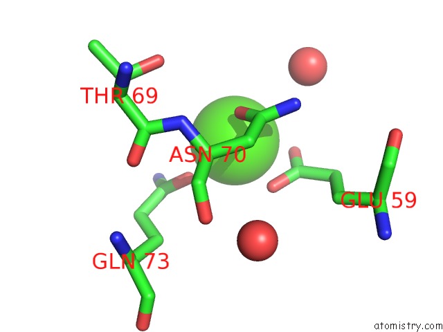
Mono view
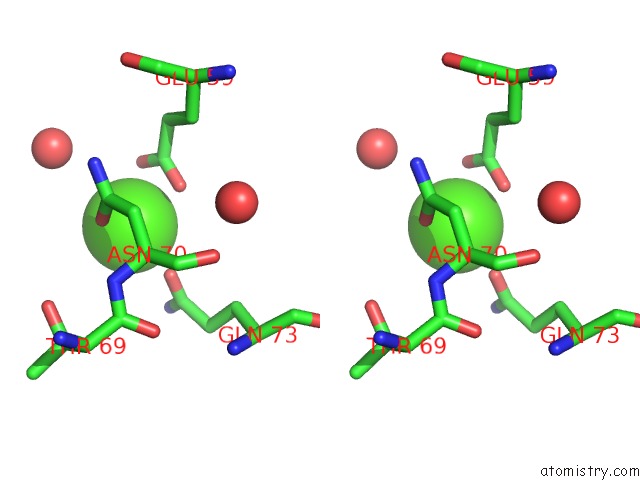
Stereo pair view
Mono view
Stereo pair view
A full contact list of Calcium with other atoms in the Ca binding
site number 2 of The Crystal Structure of the Cathepsin K Variant M5 in Complex with Chondroitin-4-Sulfate within 5.0Å range:
|
Reference:
M.M.Cherney,
F.Lecaille,
M.Kienitz,
F.S.Nallaseth,
Z.Li,
M.N.James,
D.Bromme.
Structure-Activity Analysis of Cathepsin K/Chondroitin 4-Sulfate Interactions. J.Biol.Chem. V. 286 8988 2011.
ISSN: ISSN 0021-9258
PubMed: 21193413
DOI: 10.1074/JBC.M110.126706
Page generated: Tue Jul 8 12:52:38 2025
ISSN: ISSN 0021-9258
PubMed: 21193413
DOI: 10.1074/JBC.M110.126706
Last articles
Mg in 6UWDMg in 6UV4
Mg in 6UV3
Mg in 6UV2
Mg in 6UT8
Mg in 6UT6
Mg in 6UV0
Mg in 6UT5
Mg in 6UT4
Mg in 6UT3