Calcium »
PDB 3gy6-3hii »
3hbt »
Calcium in PDB 3hbt: The Structure of Native G-Actin
Protein crystallography data
The structure of The Structure of Native G-Actin, PDB code: 3hbt
was solved by
H.Wang,
R.C.Robinson,
L.D.Burtnick,
with X-Ray Crystallography technique. A brief refinement statistics is given in the table below:
| Resolution Low / High (Å) | 27.64 / 2.70 |
| Space group | P 32 2 1 |
| Cell size a, b, c (Å), α, β, γ (°) | 95.731, 95.731, 96.948, 90.00, 90.00, 120.00 |
| R / Rfree (%) | 20.2 / 25.7 |
Calcium Binding Sites:
The binding sites of Calcium atom in the The Structure of Native G-Actin
(pdb code 3hbt). This binding sites where shown within
5.0 Angstroms radius around Calcium atom.
In total only one binding site of Calcium was determined in the The Structure of Native G-Actin, PDB code: 3hbt:
In total only one binding site of Calcium was determined in the The Structure of Native G-Actin, PDB code: 3hbt:
Calcium binding site 1 out of 1 in 3hbt
Go back to
Calcium binding site 1 out
of 1 in the The Structure of Native G-Actin
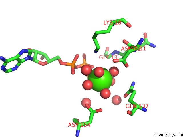
Mono view
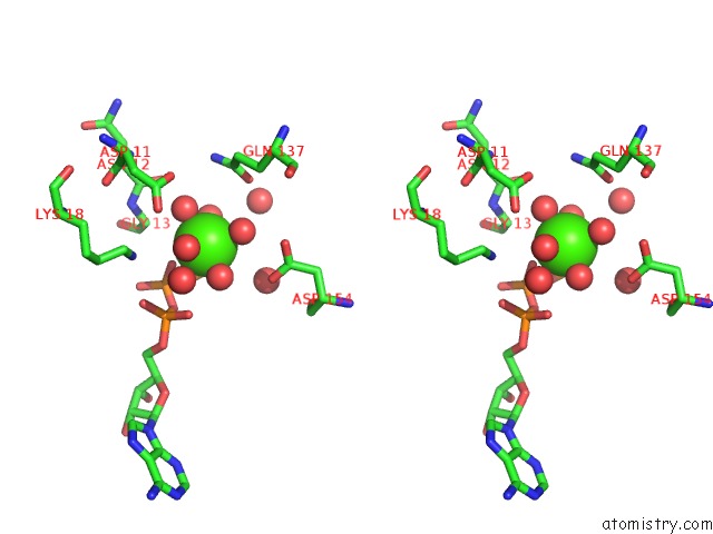
Stereo pair view
Mono view
Stereo pair view
A full contact list of Calcium with other atoms in the Ca binding
site number 1 of The Structure of Native G-Actin within 5.0Å range:
|
Reference:
H.Wang,
R.C.Robinson,
L.D.Burtnick.
The Structure of Native G-Actin Cytoskeleton (Hoboken) V. 67 456 2010.
ISSN: ISSN 1949-3584
PubMed: 20540085
DOI: 10.1002/CM.20458
Page generated: Tue Jul 8 12:54:03 2025
ISSN: ISSN 1949-3584
PubMed: 20540085
DOI: 10.1002/CM.20458
Last articles
Mg in 5SK9Mg in 5SK7
Mg in 5SK6
Mg in 5SK5
Mg in 5SK8
Mg in 5SK4
Mg in 5SK3
Mg in 5SK2
Mg in 5SK1
Mg in 5SK0