Calcium »
PDB 3hzb-3iit »
3hzx »
Calcium in PDB 3hzx: Crystal Structure of Staphylococcal Nuclease Variant D+Phs/V66K at pH 9 Determined at 100 K
Enzymatic activity of Crystal Structure of Staphylococcal Nuclease Variant D+Phs/V66K at pH 9 Determined at 100 K
All present enzymatic activity of Crystal Structure of Staphylococcal Nuclease Variant D+Phs/V66K at pH 9 Determined at 100 K:
3.1.31.1;
3.1.31.1;
Protein crystallography data
The structure of Crystal Structure of Staphylococcal Nuclease Variant D+Phs/V66K at pH 9 Determined at 100 K, PDB code: 3hzx
was solved by
J.L.Schlessman,
J.N.De Luca-Westrate,
B.E.Garcia-Moreno,
with X-Ray Crystallography technique. A brief refinement statistics is given in the table below:
| Resolution Low / High (Å) | 37.34 / 2.00 |
| Space group | P 1 21 1 |
| Cell size a, b, c (Å), α, β, γ (°) | 31.051, 60.439, 37.459, 90.00, 94.61, 90.00 |
| R / Rfree (%) | 17.8 / 23.7 |
Calcium Binding Sites:
The binding sites of Calcium atom in the Crystal Structure of Staphylococcal Nuclease Variant D+Phs/V66K at pH 9 Determined at 100 K
(pdb code 3hzx). This binding sites where shown within
5.0 Angstroms radius around Calcium atom.
In total only one binding site of Calcium was determined in the Crystal Structure of Staphylococcal Nuclease Variant D+Phs/V66K at pH 9 Determined at 100 K, PDB code: 3hzx:
In total only one binding site of Calcium was determined in the Crystal Structure of Staphylococcal Nuclease Variant D+Phs/V66K at pH 9 Determined at 100 K, PDB code: 3hzx:
Calcium binding site 1 out of 1 in 3hzx
Go back to
Calcium binding site 1 out
of 1 in the Crystal Structure of Staphylococcal Nuclease Variant D+Phs/V66K at pH 9 Determined at 100 K
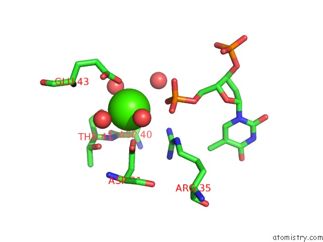
Mono view
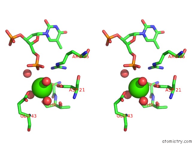
Stereo pair view
Mono view
Stereo pair view
A full contact list of Calcium with other atoms in the Ca binding
site number 1 of Crystal Structure of Staphylococcal Nuclease Variant D+Phs/V66K at pH 9 Determined at 100 K within 5.0Å range:
|
Reference:
M.J.Harms,
J.L.Schlessman,
J.N.De Luca-Westrate,
B.E.Garcia-Moreno.
Structure of Staphylococcal Nuclease D+Phs/V66K Reveals Internal Hydration in Protein Cavity To Be Published.
Page generated: Tue Jul 8 13:11:13 2025
Last articles
Mg in 4NXIMg in 4NX8
Mg in 4NV0
Mg in 4NWI
Mg in 4NX5
Mg in 4NV3
Mg in 4NW7
Mg in 4NRU
Mg in 4NST
Mg in 4NUA