Calcium »
PDB 3hzb-3iit »
3i9g »
Calcium in PDB 3i9g: Crystal Structure of the LT1009 (Sonepcizumab) Antibody Fab Fragment in Complex with Sphingosine-1-Phosphate
Protein crystallography data
The structure of Crystal Structure of the LT1009 (Sonepcizumab) Antibody Fab Fragment in Complex with Sphingosine-1-Phosphate, PDB code: 3i9g
was solved by
T.Huxford,
with X-Ray Crystallography technique. A brief refinement statistics is given in the table below:
| Resolution Low / High (Å) | 50.00 / 1.90 |
| Space group | P 21 21 21 |
| Cell size a, b, c (Å), α, β, γ (°) | 66.052, 70.889, 138.719, 90.00, 90.00, 90.00 |
| R / Rfree (%) | 19 / 21.9 |
Other elements in 3i9g:
The structure of Crystal Structure of the LT1009 (Sonepcizumab) Antibody Fab Fragment in Complex with Sphingosine-1-Phosphate also contains other interesting chemical elements:
| Magnesium | (Mg) | 5 atoms |
Calcium Binding Sites:
The binding sites of Calcium atom in the Crystal Structure of the LT1009 (Sonepcizumab) Antibody Fab Fragment in Complex with Sphingosine-1-Phosphate
(pdb code 3i9g). This binding sites where shown within
5.0 Angstroms radius around Calcium atom.
In total 2 binding sites of Calcium where determined in the Crystal Structure of the LT1009 (Sonepcizumab) Antibody Fab Fragment in Complex with Sphingosine-1-Phosphate, PDB code: 3i9g:
Jump to Calcium binding site number: 1; 2;
In total 2 binding sites of Calcium where determined in the Crystal Structure of the LT1009 (Sonepcizumab) Antibody Fab Fragment in Complex with Sphingosine-1-Phosphate, PDB code: 3i9g:
Jump to Calcium binding site number: 1; 2;
Calcium binding site 1 out of 2 in 3i9g
Go back to
Calcium binding site 1 out
of 2 in the Crystal Structure of the LT1009 (Sonepcizumab) Antibody Fab Fragment in Complex with Sphingosine-1-Phosphate
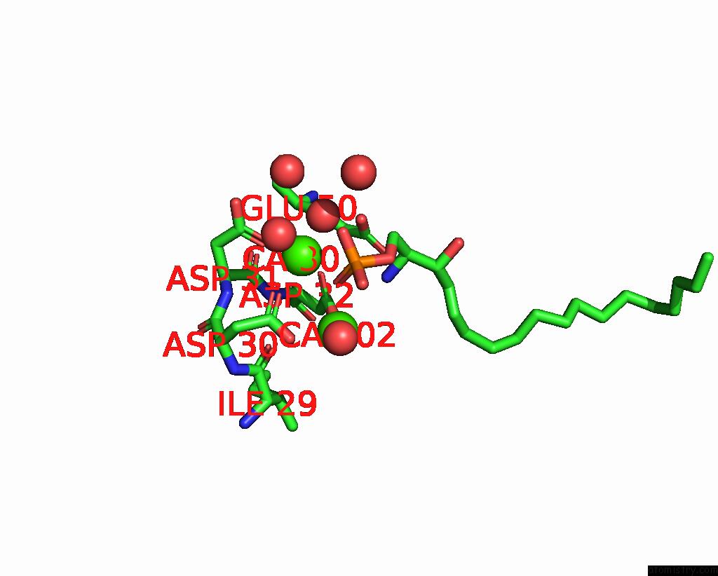
Mono view
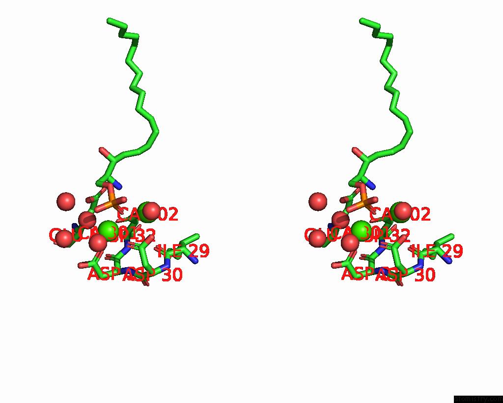
Stereo pair view
Mono view
Stereo pair view
A full contact list of Calcium with other atoms in the Ca binding
site number 1 of Crystal Structure of the LT1009 (Sonepcizumab) Antibody Fab Fragment in Complex with Sphingosine-1-Phosphate within 5.0Å range:
|
Calcium binding site 2 out of 2 in 3i9g
Go back to
Calcium binding site 2 out
of 2 in the Crystal Structure of the LT1009 (Sonepcizumab) Antibody Fab Fragment in Complex with Sphingosine-1-Phosphate
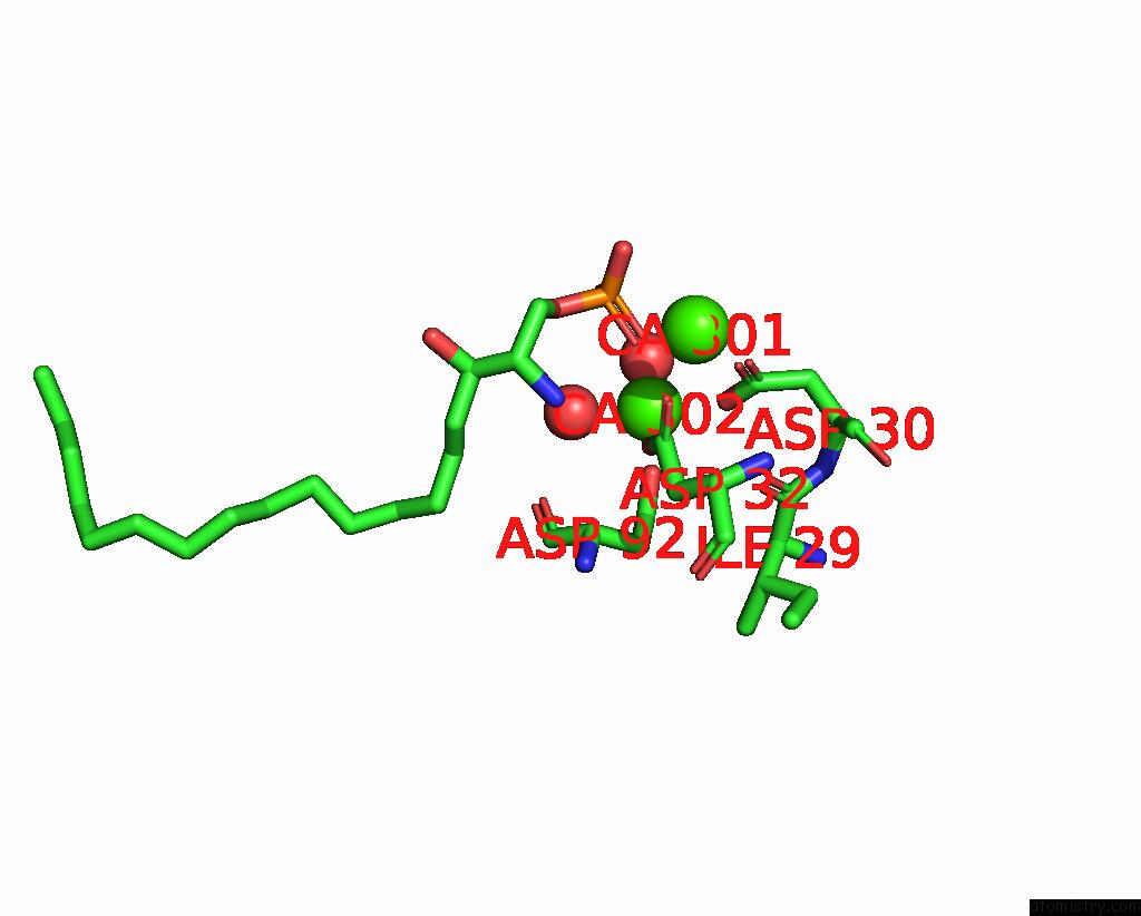
Mono view

Stereo pair view
Mono view
Stereo pair view
A full contact list of Calcium with other atoms in the Ca binding
site number 2 of Crystal Structure of the LT1009 (Sonepcizumab) Antibody Fab Fragment in Complex with Sphingosine-1-Phosphate within 5.0Å range:
|
Reference:
J.M.Wojciak,
N.Zhu,
K.T.Schuerenberg,
K.Moreno,
W.S.Shestowsky,
M.Hiraiwa,
R.Sabbadini,
T.Huxford.
The Crystal Structure of Sphingosine-1-Phosphate in Complex with A Fab Fragment Reveals Metal Bridging of An Antibody and Its Antigen. Proc.Natl.Acad.Sci.Usa V. 106 17717 2009.
ISSN: ISSN 0027-8424
PubMed: 19815502
DOI: 10.1073/PNAS.0906153106
Page generated: Tue Jul 8 13:15:57 2025
ISSN: ISSN 0027-8424
PubMed: 19815502
DOI: 10.1073/PNAS.0906153106
Last articles
Mg in 4NXIMg in 4NX8
Mg in 4NV0
Mg in 4NWI
Mg in 4NX5
Mg in 4NV3
Mg in 4NW7
Mg in 4NRU
Mg in 4NST
Mg in 4NUA