Calcium »
PDB 3ij7-3isv »
3ipk »
Calcium in PDB 3ipk: Crystal Structure of A3VP1 of Agi/II of Streptococcus Mutans
Protein crystallography data
The structure of Crystal Structure of A3VP1 of Agi/II of Streptococcus Mutans, PDB code: 3ipk
was solved by
M.R.Larson,
K.R.Rajashankar,
M.Patel,
R.Robinette,
P.Crowley,
S.M.Michalek,
L.J.Brady,
C.C.Deivanayagam,
with X-Ray Crystallography technique. A brief refinement statistics is given in the table below:
| Resolution Low / High (Å) | 42.72 / 2.04 |
| Space group | P 1 21 1 |
| Cell size a, b, c (Å), α, β, γ (°) | 50.031, 164.146, 67.733, 90.00, 91.03, 90.00 |
| R / Rfree (%) | 18.1 / 22.2 |
Calcium Binding Sites:
The binding sites of Calcium atom in the Crystal Structure of A3VP1 of Agi/II of Streptococcus Mutans
(pdb code 3ipk). This binding sites where shown within
5.0 Angstroms radius around Calcium atom.
In total 2 binding sites of Calcium where determined in the Crystal Structure of A3VP1 of Agi/II of Streptococcus Mutans, PDB code: 3ipk:
Jump to Calcium binding site number: 1; 2;
In total 2 binding sites of Calcium where determined in the Crystal Structure of A3VP1 of Agi/II of Streptococcus Mutans, PDB code: 3ipk:
Jump to Calcium binding site number: 1; 2;
Calcium binding site 1 out of 2 in 3ipk
Go back to
Calcium binding site 1 out
of 2 in the Crystal Structure of A3VP1 of Agi/II of Streptococcus Mutans
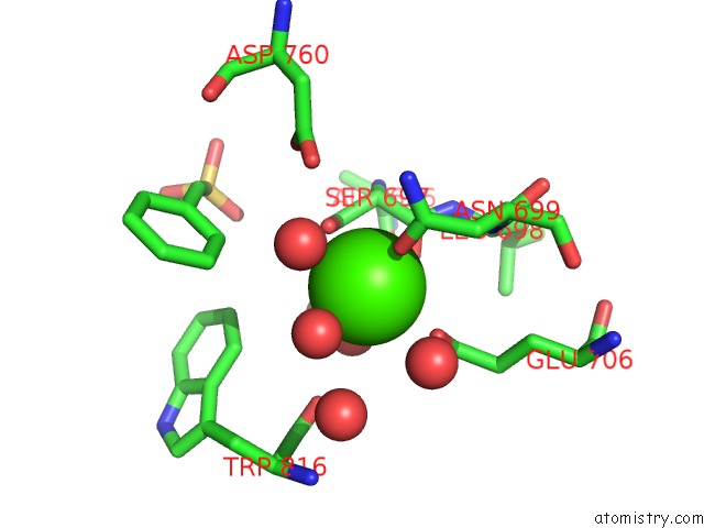
Mono view
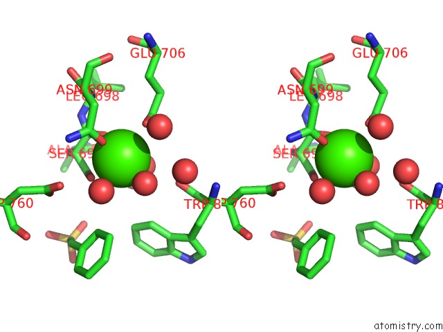
Stereo pair view
Mono view
Stereo pair view
A full contact list of Calcium with other atoms in the Ca binding
site number 1 of Crystal Structure of A3VP1 of Agi/II of Streptococcus Mutans within 5.0Å range:
|
Calcium binding site 2 out of 2 in 3ipk
Go back to
Calcium binding site 2 out
of 2 in the Crystal Structure of A3VP1 of Agi/II of Streptococcus Mutans
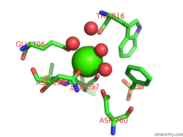
Mono view
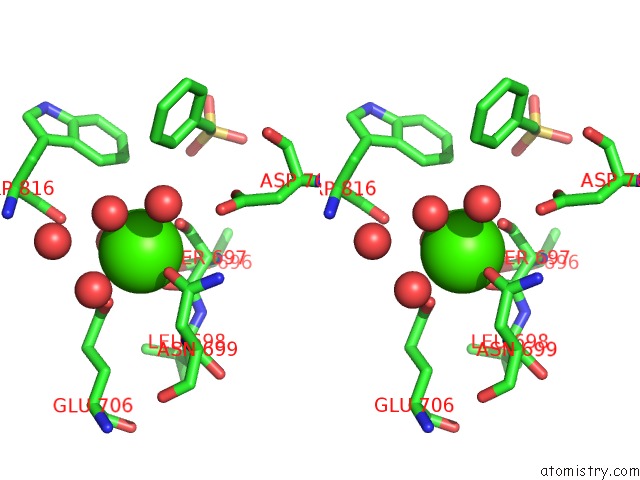
Stereo pair view
Mono view
Stereo pair view
A full contact list of Calcium with other atoms in the Ca binding
site number 2 of Crystal Structure of A3VP1 of Agi/II of Streptococcus Mutans within 5.0Å range:
|
Reference:
M.R.Larson,
K.R.Rajashankar,
M.H.Patel,
R.A.Robinette,
P.J.Crowley,
S.Michalek,
L.J.Brady,
C.Deivanayagam.
Elongated Fibrillar Structure of A Streptococcal Adhesin Assembled By the High-Affinity Association of Alpha- and Ppii-Helices. Proc.Natl.Acad.Sci.Usa V. 107 5983 2010.
ISSN: ISSN 0027-8424
PubMed: 20231452
DOI: 10.1073/PNAS.0912293107
Page generated: Tue Jul 8 13:26:52 2025
ISSN: ISSN 0027-8424
PubMed: 20231452
DOI: 10.1073/PNAS.0912293107
Last articles
Fe in 2YXOFe in 2YRS
Fe in 2YXC
Fe in 2YNM
Fe in 2YVJ
Fe in 2YP1
Fe in 2YU2
Fe in 2YU1
Fe in 2YQB
Fe in 2YOO