Calcium »
PDB 3k5m-3km5 »
3k7l »
Calcium in PDB 3k7l: Structures of Two Elapid Snake Venom Metalloproteases with Distinct Activities Highlight the Disulfide Patterns in the D Domain of Adamalysin Family Proteins
Protein crystallography data
The structure of Structures of Two Elapid Snake Venom Metalloproteases with Distinct Activities Highlight the Disulfide Patterns in the D Domain of Adamalysin Family Proteins, PDB code: 3k7l
was solved by
H.H.Guan,
W.G.Wu,
C.J.Chen,
with X-Ray Crystallography technique. A brief refinement statistics is given in the table below:
| Resolution Low / High (Å) | 30.00 / 2.50 |
| Space group | P 43 21 2 |
| Cell size a, b, c (Å), α, β, γ (°) | 91.650, 91.650, 124.235, 90.00, 90.00, 90.00 |
| R / Rfree (%) | 22.4 / 23.4 |
Other elements in 3k7l:
The structure of Structures of Two Elapid Snake Venom Metalloproteases with Distinct Activities Highlight the Disulfide Patterns in the D Domain of Adamalysin Family Proteins also contains other interesting chemical elements:
| Zinc | (Zn) | 1 atom |
Calcium Binding Sites:
The binding sites of Calcium atom in the Structures of Two Elapid Snake Venom Metalloproteases with Distinct Activities Highlight the Disulfide Patterns in the D Domain of Adamalysin Family Proteins
(pdb code 3k7l). This binding sites where shown within
5.0 Angstroms radius around Calcium atom.
In total 3 binding sites of Calcium where determined in the Structures of Two Elapid Snake Venom Metalloproteases with Distinct Activities Highlight the Disulfide Patterns in the D Domain of Adamalysin Family Proteins, PDB code: 3k7l:
Jump to Calcium binding site number: 1; 2; 3;
In total 3 binding sites of Calcium where determined in the Structures of Two Elapid Snake Venom Metalloproteases with Distinct Activities Highlight the Disulfide Patterns in the D Domain of Adamalysin Family Proteins, PDB code: 3k7l:
Jump to Calcium binding site number: 1; 2; 3;
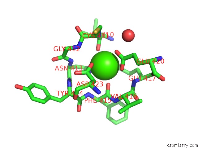
Calcium binding site 1 out of 3 in 3k7l
Go back to
Calcium binding site 1 out
of 3 in the Structures of Two Elapid Snake Venom Metalloproteases with Distinct Activities Highlight the Disulfide Patterns in the D Domain of Adamalysin Family Proteins
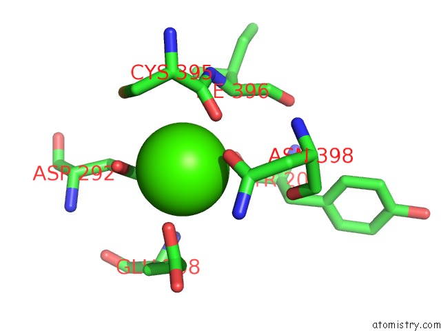
Mono view
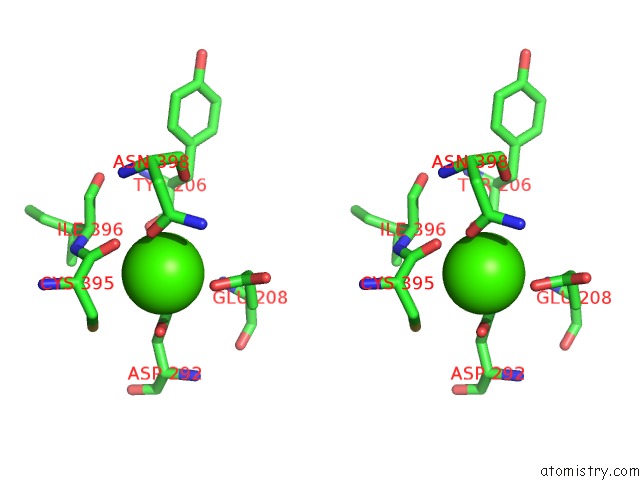
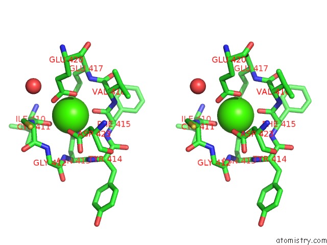
Stereo pair view
Mono view
Stereo pair view
A full contact list of Calcium with other atoms in the Ca binding
site number 1 of Structures of Two Elapid Snake Venom Metalloproteases with Distinct Activities Highlight the Disulfide Patterns in the D Domain of Adamalysin Family Proteins within 5.0Å range:
|
Calcium binding site 2 out of 3 in 3k7l
Go back to
Calcium binding site 2 out
of 3 in the Structures of Two Elapid Snake Venom Metalloproteases with Distinct Activities Highlight the Disulfide Patterns in the D Domain of Adamalysin Family Proteins
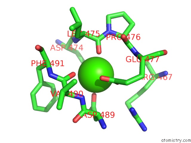
Mono view
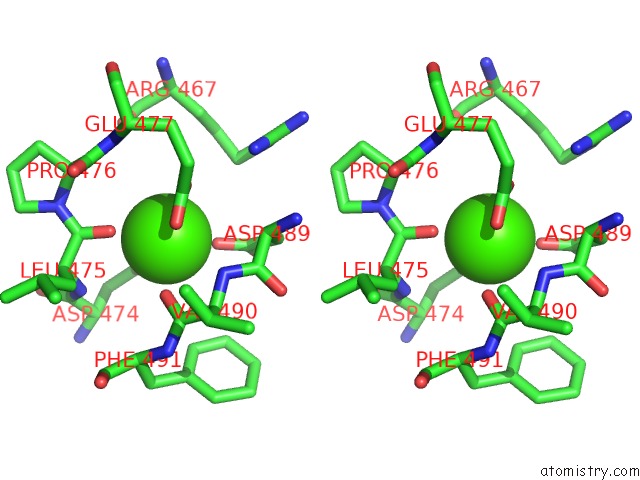
Stereo pair view
Mono view
Stereo pair view
A full contact list of Calcium with other atoms in the Ca binding
site number 2 of Structures of Two Elapid Snake Venom Metalloproteases with Distinct Activities Highlight the Disulfide Patterns in the D Domain of Adamalysin Family Proteins within 5.0Å range:
|
Calcium binding site 3 out of 3 in 3k7l
Go back to
Calcium binding site 3 out
of 3 in the Structures of Two Elapid Snake Venom Metalloproteases with Distinct Activities Highlight the Disulfide Patterns in the D Domain of Adamalysin Family Proteins
Mono view
Stereo pair view
Mono view
Stereo pair view
A full contact list of Calcium with other atoms in the Ca binding
site number 3 of Structures of Two Elapid Snake Venom Metalloproteases with Distinct Activities Highlight the Disulfide Patterns in the D Domain of Adamalysin Family Proteins within 5.0Å range:
|
Reference:
H.H.Guan,
K.S.Goh,
F.Davamani,
P.L.Wu,
Y.W.Huang,
J.Jeyakanthan,
W.G.Wu,
C.J.Chen.
Structures of Two Elapid Snake Venom Metalloproteases with Distinct Activities Highlight the Disulfide Patterns in the D Domain of Adamalysin Family Proteins J.Struct.Biol. V. 169 294 2010.
ISSN: ISSN 1047-8477
PubMed: 19932752
DOI: 10.1016/J.JSB.2009.11.009
Page generated: Tue Jul 8 13:42:51 2025
ISSN: ISSN 1047-8477
PubMed: 19932752
DOI: 10.1016/J.JSB.2009.11.009
Last articles
Mg in 7OIKMg in 7OIE
Mg in 7OI8
Mg in 7OF0
Mg in 7OI7
Mg in 7OI6
Mg in 7OGN
Mg in 7OHN
Mg in 7OHK
Mg in 7OHG