Atomistry » Calcium » PDB 3km6-3l4p » 3ko0
Atomistry »
  Calcium »
    PDB 3km6-3l4p »
      3ko0 »

Calcium in PDB 3ko0: Structure of the Tfp-CA2+-Bound Activated Form of the S100A4 Metastasis Factor

Protein crystallography data

The structure of Structure of the Tfp-CA2+-Bound Activated Form of the S100A4 Metastasis Factor, PDB code: 3ko0 was solved by V.N.Malashkevich, N.G.Dulyaninova, D.Knight, S.C.Almo, A.R.Bresnick, with X-Ray Crystallography technique. A brief refinement statistics is given in the table below:

Resolution Low / High (Å) 19.94 / 2.30
Space group P 1 21 1
Cell size a, b, c (Å), α, β, γ (°) 108.794, 102.492, 116.629, 90.00, 92.59, 90.00
R / Rfree (%) 20.6 / 25.9

Other elements in 3ko0:

The structure of Structure of the Tfp-CA2+-Bound Activated Form of the S100A4 Metastasis Factor also contains other interesting chemical elements:

Fluorine (F) 120 atoms

Calcium Binding Sites:

Pages:

>>> Page 1 <<< Page 2, Binding sites: 11 - 20; Page 3, Binding sites: 21 - 30; Page 4, Binding sites: 31 - 40;

Binding sites:

The binding sites of Calcium atom in the Structure of the Tfp-CA2+-Bound Activated Form of the S100A4 Metastasis Factor (pdb code 3ko0). This binding sites where shown within 5.0 Angstroms radius around Calcium atom.
In total 40 binding sites of Calcium where determined in the Structure of the Tfp-CA2+-Bound Activated Form of the S100A4 Metastasis Factor, PDB code: 3ko0:
Jump to Calcium binding site number: 1; 2; 3; 4; 5; 6; 7; 8; 9; 10;

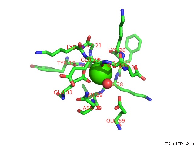
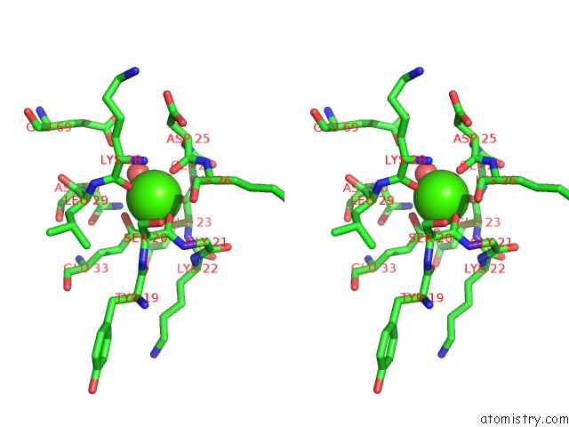
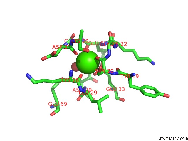
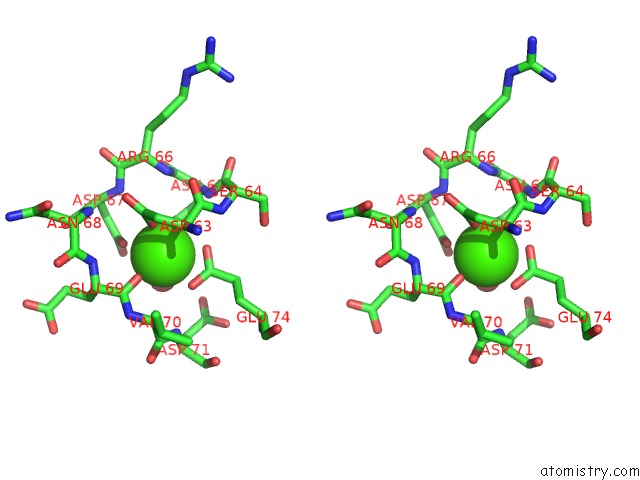
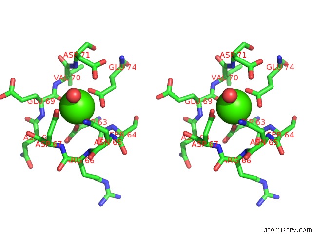
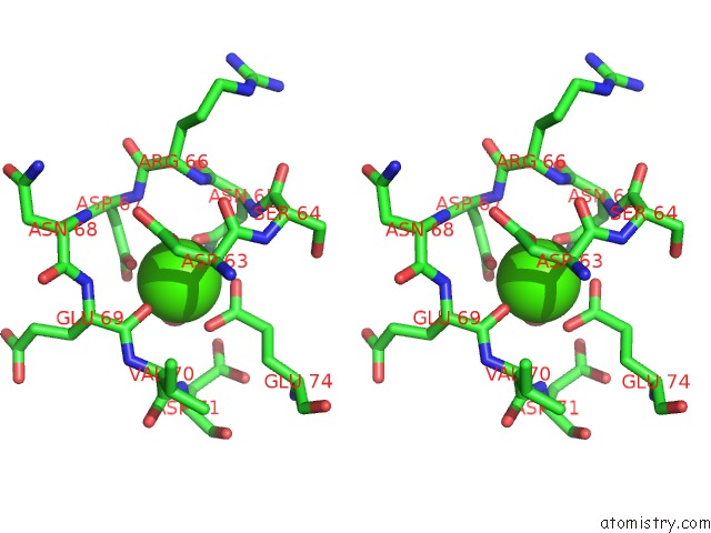
Calcium binding site 1 out of 40 in 3ko0

Go back to Calcium Binding Sites List in 3ko0
Calcium binding site 1 out of 40 in the Structure of the Tfp-CA2+-Bound Activated Form of the S100A4 Metastasis Factor


Mono view


Stereo pair view

A full contact list of Calcium with other atoms in the Ca binding site number 1 of Structure of the Tfp-CA2+-Bound Activated Form of the S100A4 Metastasis Factor within 5.0Å range:
probe atom residue distance (Å) B Occ
A:Ca301

b:15.5
occ:1.00
OE2 A:GLU33 2.2 14.3 1.0
O A:SER20 2.2 17.6 1.0
O A:ASP25 2.3 17.2 1.0
O A:LYS28 2.3 12.0 1.0
O A:HOH482 2.3 19.7 1.0
O A:GLU23 2.5 21.2 1.0
OE1 A:GLU33 2.6 13.4 1.0
CD A:GLU33 2.8 13.1 1.0
C A:SER20 3.4 17.2 1.0
C A:ASP25 3.5 17.9 1.0
C A:LYS28 3.5 13.2 1.0
C A:GLU23 3.6 20.6 1.0
CA A:SER20 4.0 15.9 1.0
N A:GLU23 4.1 21.6 1.0
N A:ASP25 4.2 18.0 1.0
N A:LYS28 4.3 14.8 1.0
CG A:GLU33 4.3 10.8 1.0
CA A:GLU23 4.4 22.4 1.0
CA A:ASP25 4.4 18.6 1.0
C A:GLY24 4.4 17.7 1.0
N A:LYS26 4.4 18.7 1.0
CA A:LYS28 4.4 14.9 1.0
N A:ASN30 4.4 17.4 1.0
N A:GLY21 4.4 18.6 1.0
N A:LEU29 4.5 12.6 1.0
CA A:LEU29 4.5 14.2 1.0
CA A:LYS26 4.5 19.4 1.0
O A:GLY24 4.6 17.6 1.0
N A:GLY24 4.7 18.5 1.0
CB A:SER20 4.7 15.0 1.0
C A:LYS26 4.7 18.5 1.0
OE2 A:GLU69 4.7 58.1 1.0
CA A:GLY21 4.7 19.1 1.0
O A:TYR19 4.7 14.1 1.0
N A:LYS22 4.8 20.8 1.0
CB A:GLU23 4.8 23.9 1.0
CB A:ASP25 4.8 20.0 1.0
C A:LEU29 4.8 16.5 1.0
CA A:GLY24 4.9 17.4 1.0
CB A:LYS28 4.9 17.9 1.0
C A:GLY21 5.0 19.6 1.0
N A:PHE27 5.0 17.8 1.0
ND2 A:ASN30 5.0 15.7 1.0

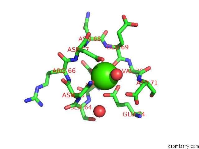
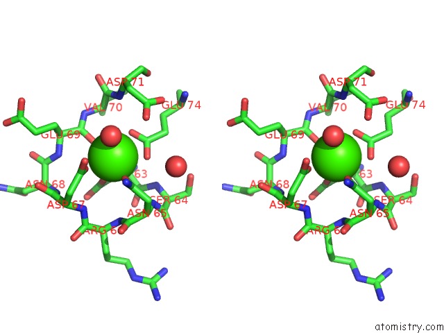
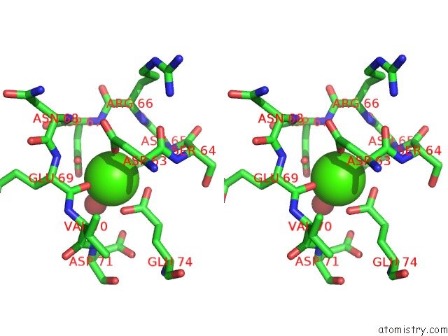
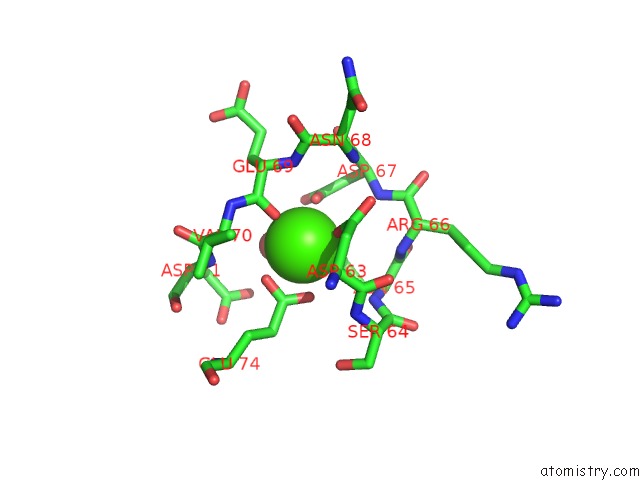
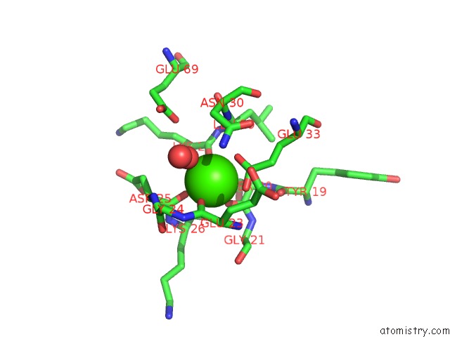
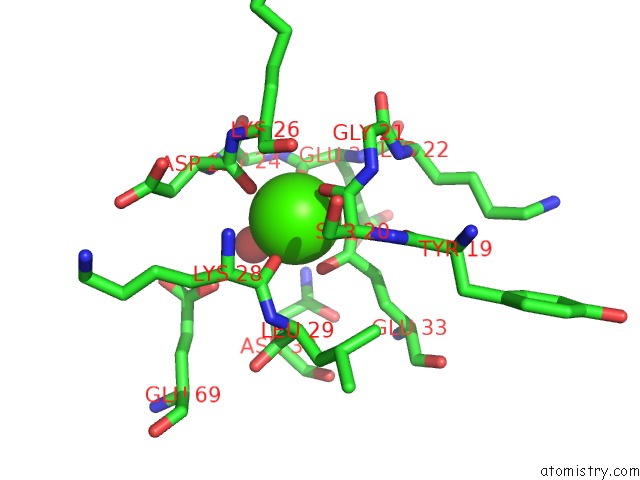
Calcium binding site 2 out of 40 in 3ko0

Go back to Calcium Binding Sites List in 3ko0
Calcium binding site 2 out of 40 in the Structure of the Tfp-CA2+-Bound Activated Form of the S100A4 Metastasis Factor


Mono view


Stereo pair view

A full contact list of Calcium with other atoms in the Ca binding site number 2 of Structure of the Tfp-CA2+-Bound Activated Form of the S100A4 Metastasis Factor within 5.0Å range:
probe atom residue distance (Å) B Occ
A:Ca302

b:12.6
occ:1.00
OD1 A:ASN65 2.2 11.7 1.0
OD1 A:ASP63 2.2 13.4 1.0
O A:GLU69 2.2 16.8 1.0
OD1 A:ASP67 2.4 13.7 1.0
O A:HOH1700 2.4 15.1 1.0
OE1 A:GLU74 2.4 12.7 1.0
OE2 A:GLU74 2.4 11.6 1.0
CD A:GLU74 2.8 17.5 1.0
CG A:ASN65 3.4 16.5 1.0
CG A:ASP67 3.4 31.4 1.0
C A:GLU69 3.4 17.1 1.0
CG A:ASP63 3.4 16.8 1.0
OD2 A:ASP67 3.7 14.4 1.0
ND2 A:ASN65 4.0 11.3 1.0
CA A:ASP63 4.1 17.0 1.0
N A:GLU69 4.1 18.1 1.0
CB A:ASP63 4.2 15.3 1.0
N A:ASP67 4.3 12.5 1.0
CG A:GLU74 4.3 10.9 1.0
N A:VAL70 4.3 14.8 1.0
N A:ASP71 4.3 11.7 1.0
CA A:GLU69 4.3 18.5 1.0
CA A:VAL70 4.3 13.6 1.0
N A:ASN65 4.4 14.7 1.0
OD2 A:ASP63 4.4 14.4 1.0
C A:ASP63 4.5 17.7 1.0
CB A:ASN65 4.5 12.1 1.0
OD2 A:ASP71 4.6 22.9 1.0
N A:ARG66 4.6 14.8 1.0
CB A:ASP67 4.7 12.7 1.0
N A:SER64 4.7 17.9 1.0
CB A:GLU69 4.7 20.9 1.0
CA A:ASN65 4.7 13.1 1.0
O A:HOH893 4.7 25.8 1.0
C A:VAL70 4.8 13.3 1.0
C A:ASN65 4.8 12.9 1.0
CA A:ASP67 4.9 12.8 1.0
N A:ASN68 4.9 14.0 1.0
CG A:ASP71 4.9 26.9 1.0

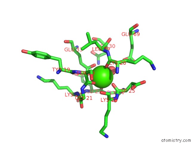
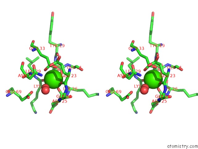
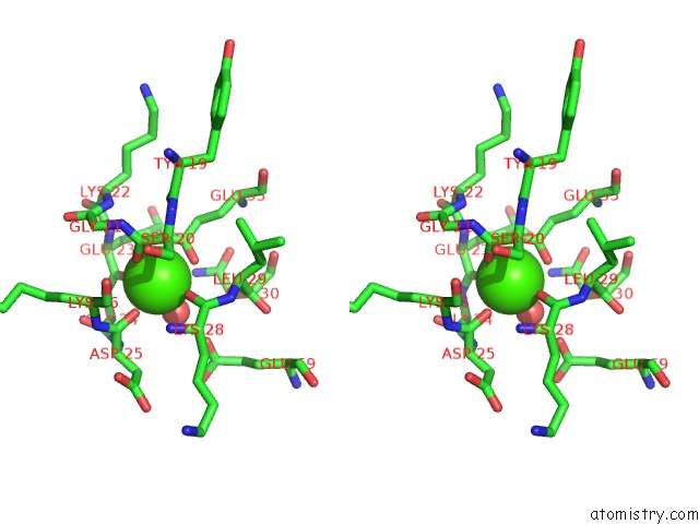
Calcium binding site 3 out of 40 in 3ko0

Go back to Calcium Binding Sites List in 3ko0
Calcium binding site 3 out of 40 in the Structure of the Tfp-CA2+-Bound Activated Form of the S100A4 Metastasis Factor


Mono view


Stereo pair view

A full contact list of Calcium with other atoms in the Ca binding site number 3 of Structure of the Tfp-CA2+-Bound Activated Form of the S100A4 Metastasis Factor within 5.0Å range:
probe atom residue distance (Å) B Occ
B:Ca301

b:14.7
occ:1.00
O B:ASP25 2.2 21.8 1.0
O B:SER20 2.3 18.7 1.0
O B:HOH162 2.3 12.9 1.0
O B:GLU23 2.3 23.5 1.0
OE1 B:GLU33 2.3 20.4 1.0
O B:LYS28 2.4 17.7 1.0
OE2 B:GLU33 2.7 21.3 1.0
CD B:GLU33 2.9 18.4 1.0
C B:SER20 3.4 19.6 1.0
C B:ASP25 3.4 22.7 1.0
C B:GLU23 3.6 25.1 1.0
C B:LYS28 3.6 19.2 1.0
CA B:SER20 4.0 19.7 1.0
N B:GLU23 4.1 27.3 1.0
N B:ASP25 4.3 23.7 1.0
N B:LYS26 4.3 23.1 1.0
N B:LYS28 4.3 21.6 1.0
C B:GLY24 4.4 24.1 1.0
CG B:GLU33 4.4 19.3 1.0
CA B:GLU23 4.4 26.6 1.0
CA B:LYS26 4.4 23.4 1.0
CA B:ASP25 4.4 23.2 1.0
N B:GLY21 4.5 20.3 1.0
N B:ASN30 4.5 18.1 1.0
N B:LEU29 4.5 18.5 1.0
OE1 B:GLU69 4.5 36.8 1.0
CA B:LEU29 4.5 16.9 1.0
CA B:LYS28 4.5 20.7 1.0
C B:LYS26 4.6 21.9 1.0
N B:GLY24 4.6 24.4 1.0
N B:LYS22 4.6 23.5 1.0
O B:GLY24 4.6 24.1 1.0
CB B:SER20 4.6 18.4 1.0
O B:TYR19 4.6 20.2 1.0
CA B:GLY24 4.8 24.3 1.0
O B:LYS26 4.8 22.0 1.0
CA B:GLY21 4.8 20.6 1.0
CB B:ASP25 4.9 24.1 1.0
C B:GLY21 4.9 22.1 1.0
CB B:GLU23 4.9 27.5 1.0
C B:LEU29 5.0 18.4 1.0
ND2 B:ASN30 5.0 18.1 1.0

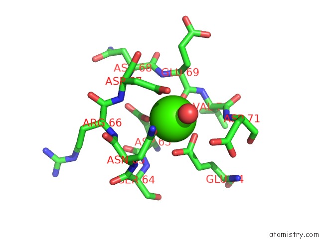
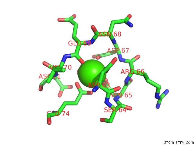
Calcium binding site 4 out of 40 in 3ko0

Go back to Calcium Binding Sites List in 3ko0
Calcium binding site 4 out of 40 in the Structure of the Tfp-CA2+-Bound Activated Form of the S100A4 Metastasis Factor


Mono view


Stereo pair view

A full contact list of Calcium with other atoms in the Ca binding site number 4 of Structure of the Tfp-CA2+-Bound Activated Form of the S100A4 Metastasis Factor within 5.0Å range:
probe atom residue distance (Å) B Occ
B:Ca302

b:18.8
occ:1.00
O B:GLU69 2.3 22.5 1.0
OD1 B:ASN65 2.3 19.9 1.0
OD1 B:ASP63 2.3 16.4 1.0
OE1 B:GLU74 2.4 20.0 1.0
OE2 B:GLU74 2.5 19.5 1.0
O B:HOH1213 2.5 17.9 1.0
OD1 B:ASP67 2.7 20.1 1.0
CD B:GLU74 2.8 14.7 1.0
CG B:ASP67 3.3 29.0 1.0
C B:GLU69 3.5 21.9 1.0
CG B:ASP63 3.5 22.2 1.0
CG B:ASN65 3.5 22.2 1.0
OD2 B:ASP67 3.6 20.6 1.0
N B:ASP67 4.0 17.6 1.0
CA B:ASP63 4.1 17.7 1.0
N B:GLU69 4.1 22.1 1.0
ND2 B:ASN65 4.2 20.7 1.0
CB B:ASP63 4.2 16.2 1.0
OD2 B:ASP63 4.3 15.9 1.0
N B:ASN65 4.3 17.7 1.0
CG B:GLU74 4.3 19.6 1.0
N B:VAL70 4.3 20.2 1.0
CA B:GLU69 4.4 22.9 1.0
N B:ARG66 4.4 16.1 1.0
CA B:VAL70 4.4 17.1 1.0
CB B:ASP67 4.4 19.4 1.0
C B:ASP63 4.5 16.6 1.0
N B:ASP71 4.5 15.8 1.0
CB B:ASN65 4.6 19.2 1.0
OD2 B:ASP71 4.6 25.1 1.0
CA B:ASP67 4.7 18.9 1.0
C B:ASN65 4.7 16.8 1.0
CA B:ASN65 4.7 18.2 1.0
N B:SER64 4.8 16.8 1.0
N B:ASN68 4.8 19.0 1.0
CG B:ASP71 4.9 34.3 1.0
C B:ASP67 4.9 19.6 1.0
C B:VAL70 4.9 15.7 1.0
O B:ASP63 5.0 16.6 1.0

Calcium binding site 5 out of 40 in 3ko0

Go back to Calcium Binding Sites List in 3ko0
Calcium binding site 5 out of 40 in the Structure of the Tfp-CA2+-Bound Activated Form of the S100A4 Metastasis Factor


Mono view


Stereo pair view

A full contact list of Calcium with other atoms in the Ca binding site number 5 of Structure of the Tfp-CA2+-Bound Activated Form of the S100A4 Metastasis Factor within 5.0Å range:
probe atom residue distance (Å) B Occ
C:Ca301

b:17.1
occ:1.00
OE2 C:GLU33 2.3 17.3 1.0
O C:LYS28 2.4 17.9 1.0
O C:SER20 2.4 15.7 1.0
O C:HOH291 2.4 23.5 1.0
O C:ASP25 2.4 20.4 1.0
O C:GLU23 2.4 27.4 1.0
OE1 C:GLU33 2.5 17.4 1.0
CD C:GLU33 2.8 18.9 1.0
C C:SER20 3.4 16.3 1.0
C C:LYS28 3.6 18.5 1.0
C C:GLU23 3.6 27.1 1.0
C C:ASP25 3.6 22.0 1.0
CA C:SER20 3.9 15.6 1.0
N C:GLU23 4.0 27.2 1.0
CA C:GLU23 4.2 27.8 1.0
CG C:GLU33 4.3 15.9 1.0
N C:LYS28 4.3 20.7 1.0
N C:ASN30 4.4 19.0 1.0
C C:GLY24 4.4 23.4 1.0
N C:ASP25 4.4 23.2 1.0
CA C:LYS28 4.5 20.3 1.0
CB C:GLU23 4.5 29.1 1.0
N C:LEU29 4.5 17.5 1.0
N C:LYS26 4.5 21.4 1.0
CA C:LYS26 4.5 20.7 1.0
CA C:LEU29 4.5 17.5 1.0
CA C:ASP25 4.6 23.5 1.0
N C:GLY21 4.6 18.4 1.0
N C:GLY24 4.6 25.6 1.0
O C:GLY24 4.6 23.2 1.0
CB C:SER20 4.7 14.3 1.0
O C:TYR19 4.7 16.7 1.0
N C:LYS22 4.7 25.0 1.0
C C:LYS26 4.7 19.6 1.0
CA C:GLY24 4.8 24.2 1.0
OE1 C:GLU69 4.8 51.8 1.0
C C:LEU29 4.9 18.5 1.0
C C:GLY21 4.9 22.4 1.0
CB C:ASP25 4.9 25.1 1.0
CA C:GLY21 5.0 19.7 1.0

Calcium binding site 6 out of 40 in 3ko0

Go back to Calcium Binding Sites List in 3ko0
Calcium binding site 6 out of 40 in the Structure of the Tfp-CA2+-Bound Activated Form of the S100A4 Metastasis Factor


Mono view


Stereo pair view

A full contact list of Calcium with other atoms in the Ca binding site number 6 of Structure of the Tfp-CA2+-Bound Activated Form of the S100A4 Metastasis Factor within 5.0Å range:
probe atom residue distance (Å) B Occ
C:Ca302

b:17.8
occ:1.00
OD1 C:ASP63 2.2 15.5 1.0
O C:HOH1841 2.2 26.1 1.0
OD1 C:ASN65 2.3 15.6 1.0
O C:GLU69 2.3 17.8 1.0
OD1 C:ASP67 2.4 19.6 1.0
OE2 C:GLU74 2.5 11.9 1.0
OE1 C:GLU74 2.5 13.1 1.0
CD C:GLU74 2.8 15.7 1.0
CG C:ASP67 3.3 31.8 1.0
CG C:ASP63 3.4 21.8 1.0
CG C:ASN65 3.4 23.1 1.0
C C:GLU69 3.4 17.9 1.0
OD2 C:ASP67 3.7 20.2 1.0
CA C:ASP63 4.0 17.3 1.0
N C:GLU69 4.1 20.6 1.0
CB C:ASP63 4.2 16.5 1.0
ND2 C:ASN65 4.2 15.8 1.0
N C:ASN65 4.2 16.2 1.0
CA C:VAL70 4.3 14.8 1.0
N C:VAL70 4.3 15.0 1.0
CA C:GLU69 4.3 19.2 1.0
CG C:GLU74 4.3 13.2 1.0
OD2 C:ASP63 4.3 16.5 1.0
N C:ASP71 4.4 14.7 1.0
N C:ASP67 4.4 14.5 1.0
CB C:ASN65 4.4 15.5 1.0
C C:ASP63 4.5 17.5 1.0
N C:SER64 4.6 16.8 1.0
CB C:ASP67 4.6 17.6 1.0
CA C:ASN65 4.6 15.7 1.0
OD2 C:ASP71 4.6 21.1 1.0
N C:ARG66 4.6 16.9 1.0
CB C:GLU69 4.7 20.3 1.0
C C:VAL70 4.8 15.3 1.0
N C:ASN68 4.8 19.9 1.0
C C:ASN65 4.8 16.1 1.0
CA C:ASP67 4.9 16.3 1.0
CG C:ASP71 5.0 43.0 1.0
C C:ASP67 5.0 18.7 1.0

Calcium binding site 7 out of 40 in 3ko0

Go back to Calcium Binding Sites List in 3ko0
Calcium binding site 7 out of 40 in the Structure of the Tfp-CA2+-Bound Activated Form of the S100A4 Metastasis Factor


Mono view


Stereo pair view

A full contact list of Calcium with other atoms in the Ca binding site number 7 of Structure of the Tfp-CA2+-Bound Activated Form of the S100A4 Metastasis Factor within 5.0Å range:
probe atom residue distance (Å) B Occ
D:Ca301

b:23.1
occ:1.00
O D:ASP25 2.2 22.9 1.0
O D:LYS28 2.4 17.8 1.0
O D:HOH1233 2.4 24.6 1.0
OE1 D:GLU33 2.4 20.7 1.0
O D:GLU23 2.4 27.7 1.0
O D:SER20 2.5 21.8 1.0
OE2 D:GLU33 2.7 21.3 1.0
CD D:GLU33 2.9 23.1 1.0
C D:ASP25 3.4 23.7 1.0
C D:SER20 3.5 22.3 1.0
C D:LYS28 3.6 18.2 1.0
C D:GLU23 3.6 27.1 1.0
CA D:SER20 4.0 20.9 1.0
N D:ASP25 4.1 25.2 1.0
N D:GLU23 4.2 26.6 1.0
CA D:ASP25 4.3 24.9 1.0
N D:LYS28 4.3 20.0 1.0
C D:GLY24 4.3 24.8 1.0
N D:LYS26 4.4 22.9 1.0
CA D:GLU23 4.4 27.4 1.0
N D:ASN30 4.4 22.3 1.0
CG D:GLU33 4.4 20.9 1.0
N D:LEU29 4.5 18.4 1.0
CA D:LYS26 4.5 23.3 1.0
CA D:LEU29 4.5 19.0 1.0
CA D:LYS28 4.5 18.7 1.0
CB D:SER20 4.6 19.3 1.0
C D:LYS26 4.6 22.5 1.0
N D:GLY24 4.6 26.1 1.0
N D:GLY21 4.7 23.2 1.0
O D:GLY24 4.7 24.7 1.0
CB D:ASP25 4.7 26.6 1.0
OE1 D:GLU69 4.7 42.1 1.0
ND2 D:ASN30 4.8 23.1 1.0
CA D:GLY24 4.8 25.0 1.0
O D:TYR19 4.8 22.5 1.0
C D:LEU29 4.8 20.8 1.0
O D:LYS26 4.9 22.8 1.0
O D:HOH273 4.9 29.6 1.0

Calcium binding site 8 out of 40 in 3ko0

Go back to Calcium Binding Sites List in 3ko0
Calcium binding site 8 out of 40 in the Structure of the Tfp-CA2+-Bound Activated Form of the S100A4 Metastasis Factor


Mono view


Stereo pair view

A full contact list of Calcium with other atoms in the Ca binding site number 8 of Structure of the Tfp-CA2+-Bound Activated Form of the S100A4 Metastasis Factor within 5.0Å range:
probe atom residue distance (Å) B Occ
D:Ca302

b:19.9
occ:1.00
O D:GLU69 2.3 19.6 1.0
O D:HOH230 2.3 23.6 1.0
OD1 D:ASN65 2.3 22.6 1.0
OE1 D:GLU74 2.3 18.4 1.0
OD1 D:ASP67 2.5 19.1 1.0
OD1 D:ASP63 2.5 20.9 1.0
OE2 D:GLU74 2.6 17.6 1.0
CD D:GLU74 2.8 20.5 1.0
CG D:ASP67 3.3 27.9 1.0
C D:GLU69 3.4 19.8 1.0
CG D:ASN65 3.5 20.7 1.0
CG D:ASP63 3.6 23.0 1.0
OD2 D:ASP67 3.7 19.3 1.0
N D:GLU69 4.0 20.9 1.0
OD1 D:ASP71 4.0 23.7 1.0
ND2 D:ASN65 4.1 22.9 1.0
N D:ASP71 4.2 17.4 1.0
CA D:GLU69 4.2 20.8 1.0
N D:VAL70 4.2 19.1 1.0
CA D:VAL70 4.2 18.6 1.0
N D:ASP67 4.3 21.7 1.0
CA D:ASP63 4.3 23.0 1.0
CG D:GLU74 4.4 18.0 1.0
CB D:ASP63 4.4 21.7 1.0
CB D:GLU69 4.4 20.6 1.0
N D:ASN65 4.5 22.3 1.0
OD2 D:ASP63 4.6 20.8 1.0
CB D:ASP67 4.6 19.6 1.0
CG D:ASP71 4.6 30.8 1.0
N D:ARG66 4.6 23.3 1.0
CB D:ASN65 4.6 22.7 1.0
C D:ASP63 4.6 23.8 1.0
C D:VAL70 4.6 17.4 1.0
N D:SER64 4.7 23.4 1.0
CA D:ASP67 4.8 20.6 1.0
N D:ASN68 4.9 21.4 1.0
CA D:ASN65 4.9 22.8 1.0
C D:ASP67 5.0 21.1 1.0
C D:ASN65 5.0 22.8 1.0

Calcium binding site 9 out of 40 in 3ko0

Go back to Calcium Binding Sites List in 3ko0
Calcium binding site 9 out of 40 in the Structure of the Tfp-CA2+-Bound Activated Form of the S100A4 Metastasis Factor


Mono view


Stereo pair view

A full contact list of Calcium with other atoms in the Ca binding site number 9 of Structure of the Tfp-CA2+-Bound Activated Form of the S100A4 Metastasis Factor within 5.0Å range:
probe atom residue distance (Å) B Occ
E:Ca301

b:19.6
occ:1.00
O E:ASP25 2.2 20.0 1.0
OE1 E:GLU33 2.3 19.6 1.0
O E:GLU23 2.3 24.1 1.0
O E:SER20 2.3 17.0 1.0
O E:LYS28 2.4 21.2 1.0
OE2 E:GLU33 2.5 18.9 1.0
O E:HOH153 2.5 14.6 1.0
CD E:GLU33 2.7 14.3 1.0
C E:SER20 3.4 18.0 1.0
C E:ASP25 3.4 21.3 1.0
C E:GLU23 3.5 23.9 1.0
C E:LYS28 3.6 20.5 1.0
CA E:SER20 3.9 18.7 1.0
N E:ASP25 4.1 21.4 1.0
CG E:GLU33 4.1 18.9 1.0
N E:GLU23 4.1 24.0 1.0
OE1 E:GLU69 4.2 39.2 1.0
C E:GLY24 4.2 21.7 1.0
N E:LYS26 4.3 21.6 1.0
CA E:LYS26 4.3 22.4 1.0
CA E:ASP25 4.4 22.2 1.0
CA E:GLU23 4.5 24.4 1.0
N E:LYS28 4.5 21.3 1.0
CA E:LEU29 4.5 17.3 1.0
N E:GLY24 4.5 23.1 1.0
N E:ASN30 4.5 20.5 1.0
O E:TYR19 4.5 18.7 1.0
N E:GLY21 4.5 18.2 1.0
N E:LEU29 4.5 18.1 1.0
CA E:GLY24 4.5 22.4 1.0
CA E:LYS28 4.6 21.8 1.0
O E:GLY24 4.6 20.6 1.0
ND2 E:ASN30 4.7 21.6 1.0
CB E:SER20 4.7 19.1 1.0
C E:LYS26 4.7 21.1 1.0
N E:LYS22 4.7 21.4 1.0
C E:LEU29 4.9 19.8 1.0
CA E:GLY21 4.9 19.1 1.0
CB E:GLU33 4.9 15.9 1.0
CB E:ASP25 4.9 24.2 1.0
CG E:GLU23 5.0 52.0 1.0
C E:GLY21 5.0 19.7 1.0

Calcium binding site 10 out of 40 in 3ko0

Go back to Calcium Binding Sites List in 3ko0
Calcium binding site 10 out of 40 in the Structure of the Tfp-CA2+-Bound Activated Form of the S100A4 Metastasis Factor


Mono view


Stereo pair view

A full contact list of Calcium with other atoms in the Ca binding site number 10 of Structure of the Tfp-CA2+-Bound Activated Form of the S100A4 Metastasis Factor within 5.0Å range:
probe atom residue distance (Å) B Occ
E:Ca302

b:23.4
occ:1.00
O E:HOH1217 2.3 27.1 1.0
OD1 E:ASP67 2.3 22.6 1.0
OD1 E:ASP63 2.3 21.7 1.0
OE1 E:GLU74 2.4 20.5 1.0
O E:GLU69 2.4 21.6 1.0
OE2 E:GLU74 2.4 22.0 1.0
OD1 E:ASN65 2.4 19.8 1.0
CD E:GLU74 2.7 17.5 1.0
CG E:ASP67 3.2 18.8 1.0
CG E:ASP63 3.5 13.3 1.0
CG E:ASN65 3.5 18.5 1.0
C E:GLU69 3.5 21.5 1.0
OD2 E:ASP67 3.7 22.9 1.0
CA E:ASP63 4.0 22.6 1.0
N E:ASP67 4.1 25.9 1.0
ND2 E:ASN65 4.1 19.6 1.0
CB E:ASP63 4.1 22.2 1.0
N E:GLU69 4.2 22.3 1.0
CG E:GLU74 4.3 21.1 1.0
N E:ASN65 4.3 20.6 1.0
C E:ASP63 4.3 21.9 1.0
CB E:ASP67 4.4 23.2 1.0
OD2 E:ASP63 4.4 22.2 1.0
N E:ARG66 4.4 25.0 1.0
N E:VAL70 4.4 20.9 1.0
CA E:VAL70 4.4 20.4 1.0
CA E:GLU69 4.5 22.5 1.0
OD2 E:ASP71 4.5 23.3 1.0
N E:ASP71 4.5 21.1 1.0
CB E:ASN65 4.6 20.3 1.0
CA E:ASP67 4.6 24.2 1.0
C E:ASN65 4.7 23.3 1.0
CA E:ASN65 4.7 21.4 1.0
N E:SER64 4.7 21.3 1.0
O E:ASP63 4.8 21.2 1.0
N E:ASN68 4.9 21.8 1.0
CG E:ASP71 4.9 30.6 1.0
C E:ASP67 4.9 23.1 1.0
C E:VAL70 5.0 20.6 1.0

Reference:

V.N.Malashkevich, N.G.Dulyaninova, U.A.Ramagopal, M.A.Liriano, K.M.Varney, D.Knight, M.Brenowitz, D.J.Weber, S.C.Almo, A.R.Bresnick. Phenothiazines Inhibit S100A4 Function By Inducing Protein Oligomerization. Proc.Natl.Acad.Sci.Usa V. 107 8605 2010.
ISSN: ISSN 0027-8424
PubMed: 20421509
DOI: 10.1073/PNAS.0913660107
Page generated: Sat Jul 13 12:33:47 2024

Last articles

Br in 4IVH
Br in 4IO1
Br in 4ILZ
Br in 4IDT
Br in 4IBF
Br in 4IBE
Br in 4IBD
Br in 4ICH
Br in 4I7T
Br in 4I7P
© Copyright 2008-2020 by atomistry.com
Home   |    Site Map   |    Copyright   |    Contact us   |    Privacy