Calcium »
PDB 3km6-3l4p »
3kqf »
Calcium in PDB 3kqf: 1.8 Angstrom Resolution Crystal Structure of Enoyl-Coa Hydratase From Bacillus Anthracis.
Enzymatic activity of 1.8 Angstrom Resolution Crystal Structure of Enoyl-Coa Hydratase From Bacillus Anthracis.
All present enzymatic activity of 1.8 Angstrom Resolution Crystal Structure of Enoyl-Coa Hydratase From Bacillus Anthracis.:
4.2.1.17;
4.2.1.17;
Protein crystallography data
The structure of 1.8 Angstrom Resolution Crystal Structure of Enoyl-Coa Hydratase From Bacillus Anthracis., PDB code: 3kqf
was solved by
G.Minasov,
A.Halavaty,
Z.Wawrzak,
T.Skarina,
O.Onopriyenko,
L.Papazisi,
A.Savchenko,
W.F.Anderson,
Center For Structural Genomics Ofinfectious Diseases (Csgid),
with X-Ray Crystallography technique. A brief refinement statistics is given in the table below:
| Resolution Low / High (Å) | 29.87 / 1.80 |
| Space group | P 1 21 1 |
| Cell size a, b, c (Å), α, β, γ (°) | 73.747, 130.721, 73.802, 90.00, 114.47, 90.00 |
| R / Rfree (%) | 15.6 / 19.3 |
Other elements in 3kqf:
The structure of 1.8 Angstrom Resolution Crystal Structure of Enoyl-Coa Hydratase From Bacillus Anthracis. also contains other interesting chemical elements:
| Chlorine | (Cl) | 2 atoms |
Calcium Binding Sites:
The binding sites of Calcium atom in the 1.8 Angstrom Resolution Crystal Structure of Enoyl-Coa Hydratase From Bacillus Anthracis.
(pdb code 3kqf). This binding sites where shown within
5.0 Angstroms radius around Calcium atom.
In total 5 binding sites of Calcium where determined in the 1.8 Angstrom Resolution Crystal Structure of Enoyl-Coa Hydratase From Bacillus Anthracis., PDB code: 3kqf:
Jump to Calcium binding site number: 1; 2; 3; 4; 5;
In total 5 binding sites of Calcium where determined in the 1.8 Angstrom Resolution Crystal Structure of Enoyl-Coa Hydratase From Bacillus Anthracis., PDB code: 3kqf:
Jump to Calcium binding site number: 1; 2; 3; 4; 5;
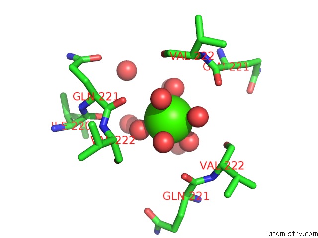
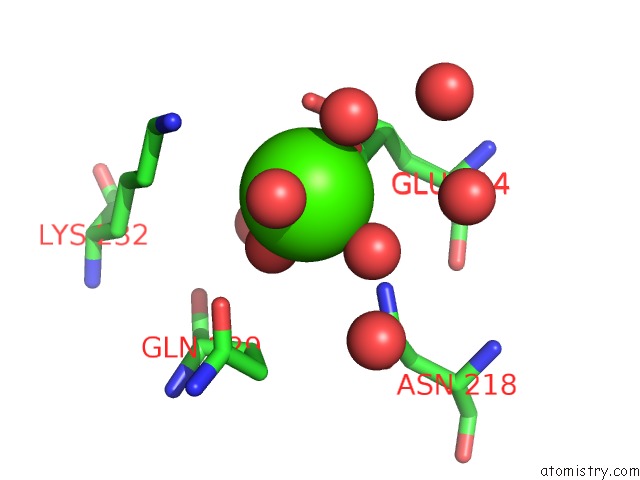
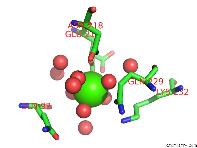
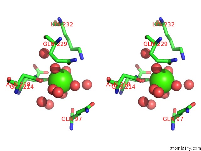
Calcium binding site 1 out of 5 in 3kqf
Go back to
Calcium binding site 1 out
of 5 in the 1.8 Angstrom Resolution Crystal Structure of Enoyl-Coa Hydratase From Bacillus Anthracis.
Mono view
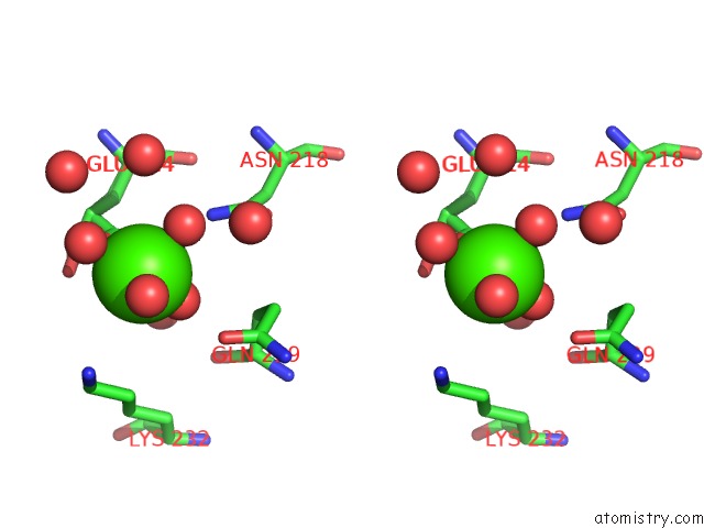
Stereo pair view
Mono view
Stereo pair view
A full contact list of Calcium with other atoms in the Ca binding
site number 1 of 1.8 Angstrom Resolution Crystal Structure of Enoyl-Coa Hydratase From Bacillus Anthracis. within 5.0Å range:
|
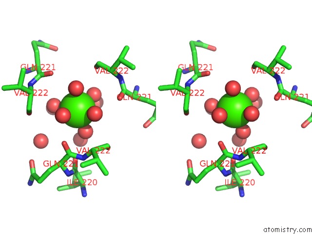
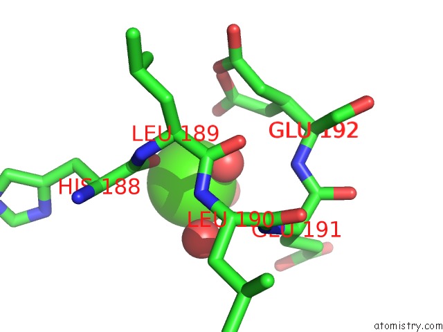
Calcium binding site 2 out of 5 in 3kqf
Go back to
Calcium binding site 2 out
of 5 in the 1.8 Angstrom Resolution Crystal Structure of Enoyl-Coa Hydratase From Bacillus Anthracis.
Mono view
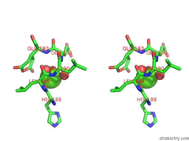
Stereo pair view
Mono view
Stereo pair view
A full contact list of Calcium with other atoms in the Ca binding
site number 2 of 1.8 Angstrom Resolution Crystal Structure of Enoyl-Coa Hydratase From Bacillus Anthracis. within 5.0Å range:
|
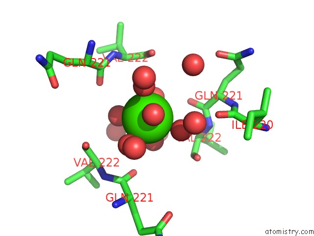
Calcium binding site 3 out of 5 in 3kqf
Go back to
Calcium binding site 3 out
of 5 in the 1.8 Angstrom Resolution Crystal Structure of Enoyl-Coa Hydratase From Bacillus Anthracis.
Mono view
Stereo pair view
Mono view
Stereo pair view
A full contact list of Calcium with other atoms in the Ca binding
site number 3 of 1.8 Angstrom Resolution Crystal Structure of Enoyl-Coa Hydratase From Bacillus Anthracis. within 5.0Å range:
|
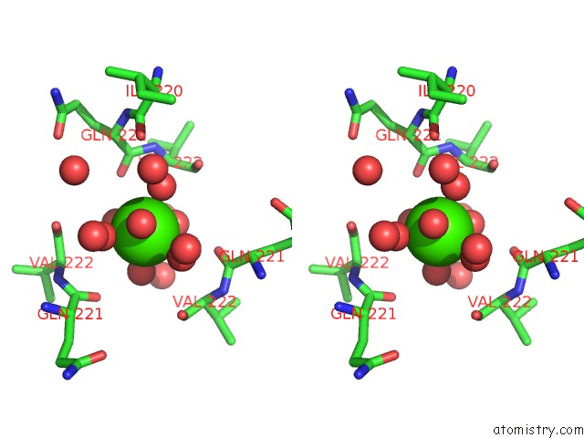
Calcium binding site 4 out of 5 in 3kqf
Go back to
Calcium binding site 4 out
of 5 in the 1.8 Angstrom Resolution Crystal Structure of Enoyl-Coa Hydratase From Bacillus Anthracis.
Mono view
Stereo pair view
Mono view
Stereo pair view
A full contact list of Calcium with other atoms in the Ca binding
site number 4 of 1.8 Angstrom Resolution Crystal Structure of Enoyl-Coa Hydratase From Bacillus Anthracis. within 5.0Å range:
|
Calcium binding site 5 out of 5 in 3kqf
Go back to
Calcium binding site 5 out
of 5 in the 1.8 Angstrom Resolution Crystal Structure of Enoyl-Coa Hydratase From Bacillus Anthracis.
Mono view
Stereo pair view
Mono view
Stereo pair view
A full contact list of Calcium with other atoms in the Ca binding
site number 5 of 1.8 Angstrom Resolution Crystal Structure of Enoyl-Coa Hydratase From Bacillus Anthracis. within 5.0Å range:
|
Reference:
G.Minasov,
A.Halavaty,
Z.Wawrzak,
T.Skarina,
O.Onopriyenko,
L.Papazisi,
A.Savchenko,
W.F.Anderson.
1.8 Angstrom Resolution Crystal Structure of Enoyl-Coa Hydratase From Bacillus Anthracis. To Be Published.
Page generated: Tue Jul 8 13:55:44 2025
Last articles
Mg in 4DPGMg in 4DQP
Mg in 4DQQ
Mg in 4DPM
Mg in 4DPV
Mg in 4DQI
Mg in 4DOB
Mg in 4DOC
Mg in 4DMZ
Mg in 4DOA