Calcium »
PDB 3l7f-3ljt »
3lcp »
Calcium in PDB 3lcp: Crystal Structure of the Carbohydrate Recognition Domain of LMAN1 in Complex with MCFD2
Protein crystallography data
The structure of Crystal Structure of the Carbohydrate Recognition Domain of LMAN1 in Complex with MCFD2, PDB code: 3lcp
was solved by
E.Wigren,
J.M.Bourhis,
I.Kursula,
J.E.Guy,
Y.Lindqvist,
with X-Ray Crystallography technique. A brief refinement statistics is given in the table below:
| Resolution Low / High (Å) | 50.00 / 2.45 |
| Space group | P 61 |
| Cell size a, b, c (Å), α, β, γ (°) | 58.602, 58.602, 396.870, 90.00, 90.00, 120.00 |
| R / Rfree (%) | 19.6 / 24.7 |
Calcium Binding Sites:
The binding sites of Calcium atom in the Crystal Structure of the Carbohydrate Recognition Domain of LMAN1 in Complex with MCFD2
(pdb code 3lcp). This binding sites where shown within
5.0 Angstroms radius around Calcium atom.
In total 8 binding sites of Calcium where determined in the Crystal Structure of the Carbohydrate Recognition Domain of LMAN1 in Complex with MCFD2, PDB code: 3lcp:
Jump to Calcium binding site number: 1; 2; 3; 4; 5; 6; 7; 8;
In total 8 binding sites of Calcium where determined in the Crystal Structure of the Carbohydrate Recognition Domain of LMAN1 in Complex with MCFD2, PDB code: 3lcp:
Jump to Calcium binding site number: 1; 2; 3; 4; 5; 6; 7; 8;
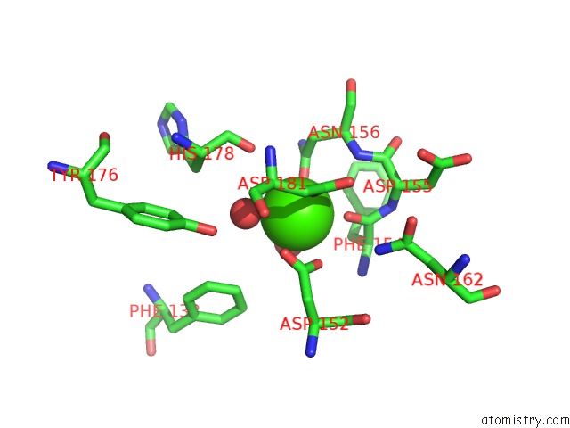
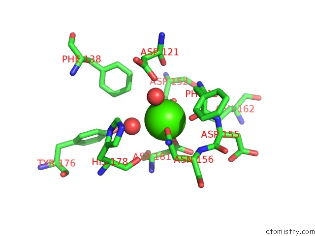
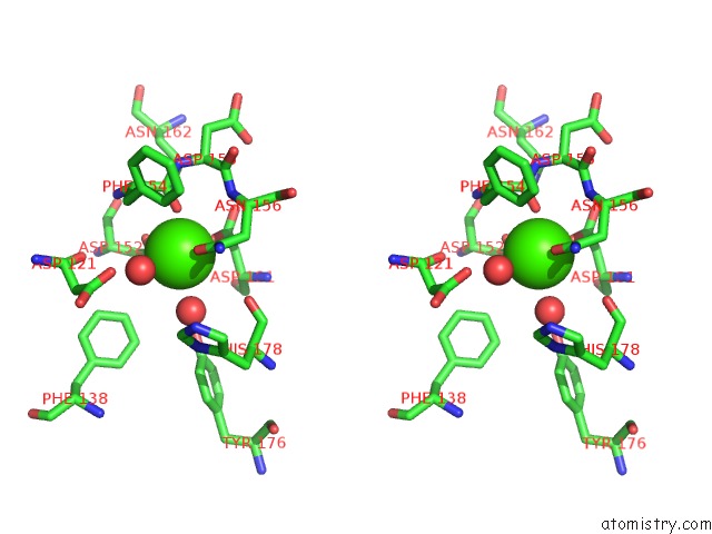
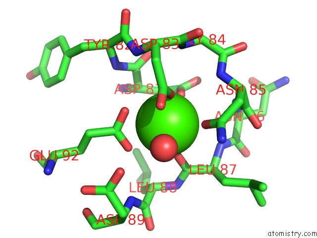
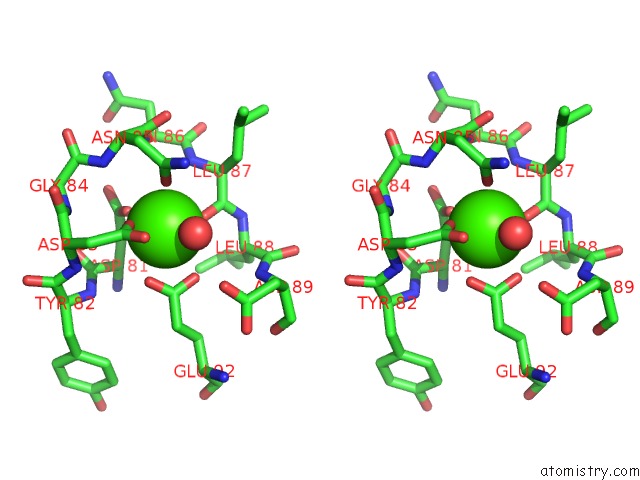
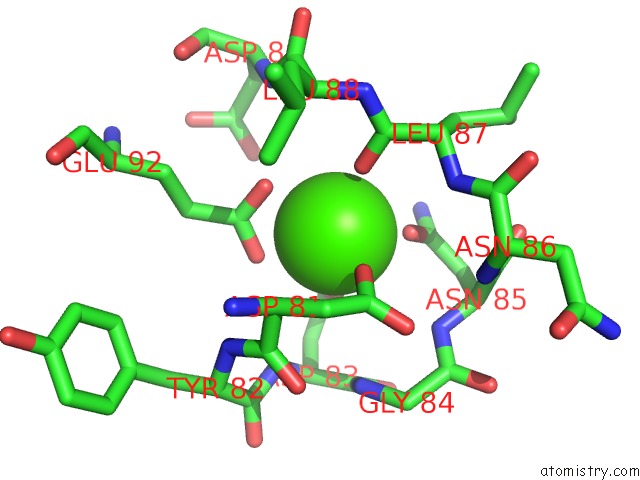
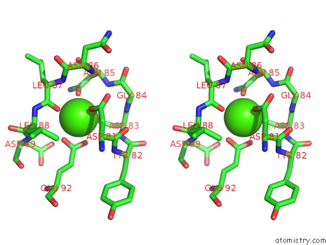
Calcium binding site 1 out of 8 in 3lcp
Go back to
Calcium binding site 1 out
of 8 in the Crystal Structure of the Carbohydrate Recognition Domain of LMAN1 in Complex with MCFD2
Mono view
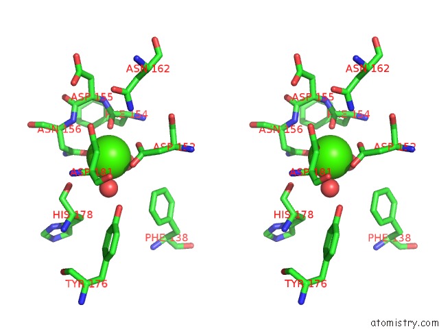
Stereo pair view
Mono view
Stereo pair view
A full contact list of Calcium with other atoms in the Ca binding
site number 1 of Crystal Structure of the Carbohydrate Recognition Domain of LMAN1 in Complex with MCFD2 within 5.0Å range:
|
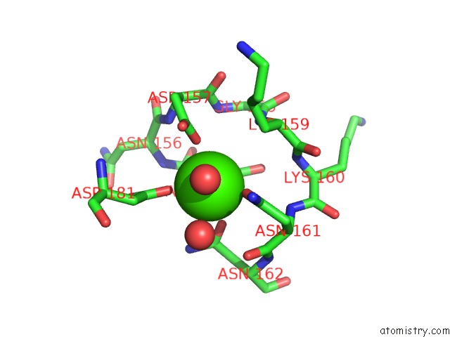
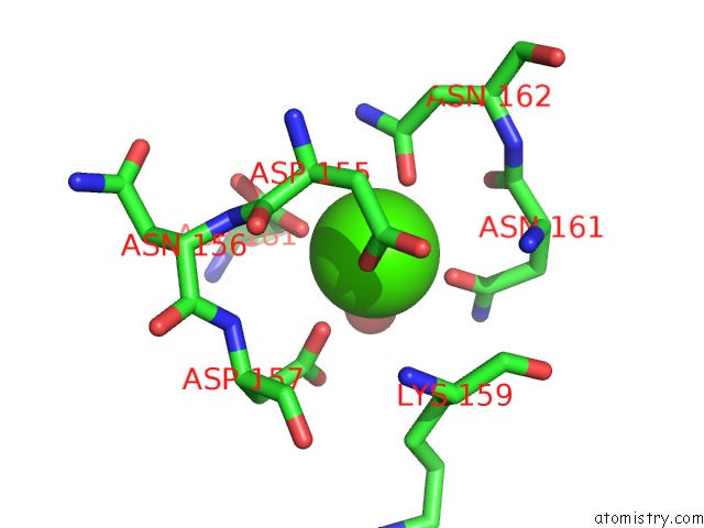
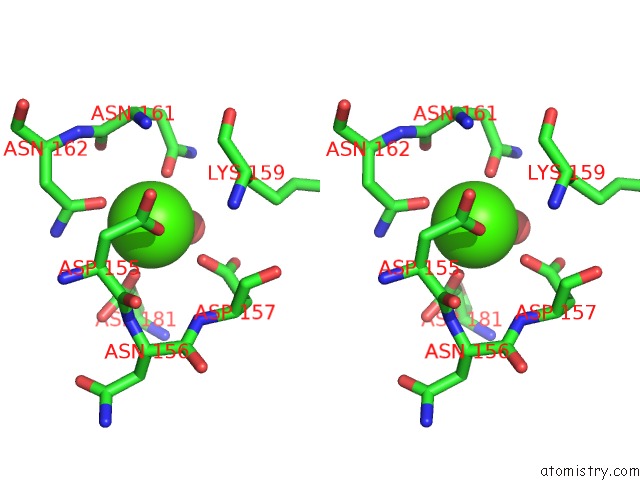
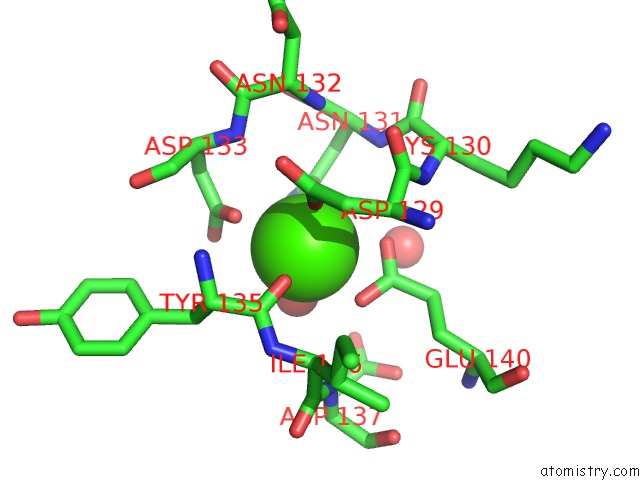
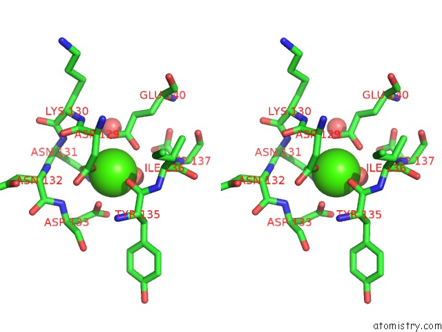
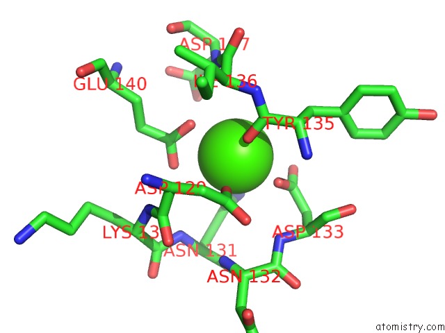
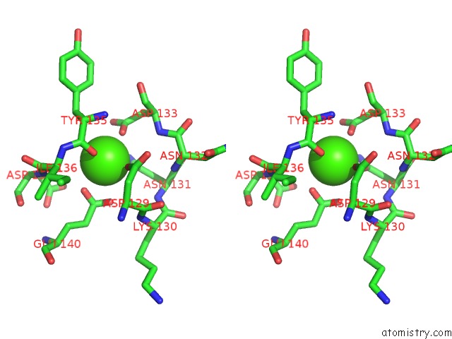
Calcium binding site 2 out of 8 in 3lcp
Go back to
Calcium binding site 2 out
of 8 in the Crystal Structure of the Carbohydrate Recognition Domain of LMAN1 in Complex with MCFD2
Mono view
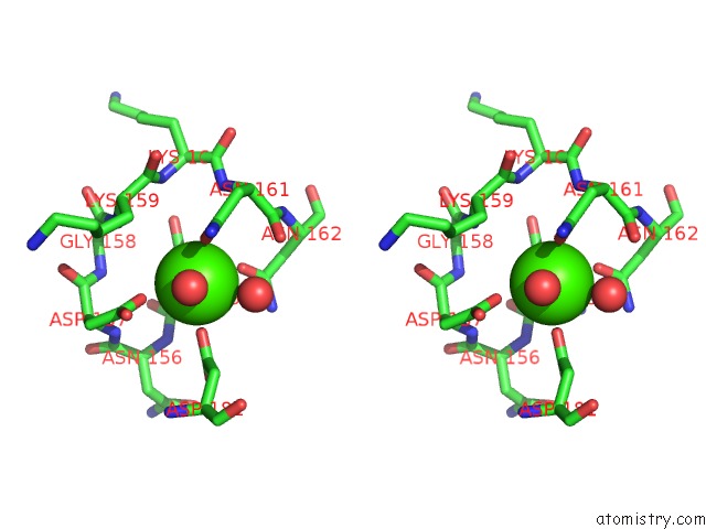
Stereo pair view
Mono view
Stereo pair view
A full contact list of Calcium with other atoms in the Ca binding
site number 2 of Crystal Structure of the Carbohydrate Recognition Domain of LMAN1 in Complex with MCFD2 within 5.0Å range:
|
Calcium binding site 3 out of 8 in 3lcp
Go back to
Calcium binding site 3 out
of 8 in the Crystal Structure of the Carbohydrate Recognition Domain of LMAN1 in Complex with MCFD2
Mono view
Stereo pair view
Mono view
Stereo pair view
A full contact list of Calcium with other atoms in the Ca binding
site number 3 of Crystal Structure of the Carbohydrate Recognition Domain of LMAN1 in Complex with MCFD2 within 5.0Å range:
|
Calcium binding site 4 out of 8 in 3lcp
Go back to
Calcium binding site 4 out
of 8 in the Crystal Structure of the Carbohydrate Recognition Domain of LMAN1 in Complex with MCFD2
Mono view
Stereo pair view
Mono view
Stereo pair view
A full contact list of Calcium with other atoms in the Ca binding
site number 4 of Crystal Structure of the Carbohydrate Recognition Domain of LMAN1 in Complex with MCFD2 within 5.0Å range:
|
Calcium binding site 5 out of 8 in 3lcp
Go back to
Calcium binding site 5 out
of 8 in the Crystal Structure of the Carbohydrate Recognition Domain of LMAN1 in Complex with MCFD2
Mono view
Stereo pair view
Mono view
Stereo pair view
A full contact list of Calcium with other atoms in the Ca binding
site number 5 of Crystal Structure of the Carbohydrate Recognition Domain of LMAN1 in Complex with MCFD2 within 5.0Å range:
|
Calcium binding site 6 out of 8 in 3lcp
Go back to
Calcium binding site 6 out
of 8 in the Crystal Structure of the Carbohydrate Recognition Domain of LMAN1 in Complex with MCFD2
Mono view
Stereo pair view
Mono view
Stereo pair view
A full contact list of Calcium with other atoms in the Ca binding
site number 6 of Crystal Structure of the Carbohydrate Recognition Domain of LMAN1 in Complex with MCFD2 within 5.0Å range:
|
Calcium binding site 7 out of 8 in 3lcp
Go back to
Calcium binding site 7 out
of 8 in the Crystal Structure of the Carbohydrate Recognition Domain of LMAN1 in Complex with MCFD2
Mono view
Stereo pair view
Mono view
Stereo pair view
A full contact list of Calcium with other atoms in the Ca binding
site number 7 of Crystal Structure of the Carbohydrate Recognition Domain of LMAN1 in Complex with MCFD2 within 5.0Å range:
|
Calcium binding site 8 out of 8 in 3lcp
Go back to
Calcium binding site 8 out
of 8 in the Crystal Structure of the Carbohydrate Recognition Domain of LMAN1 in Complex with MCFD2
Mono view
Stereo pair view
Mono view
Stereo pair view
A full contact list of Calcium with other atoms in the Ca binding
site number 8 of Crystal Structure of the Carbohydrate Recognition Domain of LMAN1 in Complex with MCFD2 within 5.0Å range:
|
Reference:
E.Wigren,
J.M.Bourhis,
I.Kursula,
J.E.Guy,
Y.Lindqvist.
Crystal Structure of the LMAN1-Crd/MCFD2 Transport Receptor Complex Provides Insight Into Combined Deficiency of Factor V and Factor VIII. Febs Lett. V. 584 878 2010.
ISSN: ISSN 0014-5793
PubMed: 20138881
DOI: 10.1016/J.FEBSLET.2010.02.009
Page generated: Tue Jul 8 14:10:40 2025
ISSN: ISSN 0014-5793
PubMed: 20138881
DOI: 10.1016/J.FEBSLET.2010.02.009
Last articles
Mg in 5SE0Mg in 5SDZ
Mg in 5SE1
Mg in 5SDX
Mg in 5SDT
Mg in 5SDY
Mg in 5SDW
Mg in 5SDV
Mg in 5SDU
Mg in 5SCL