Calcium »
PDB 3ljz-3lzk »
3lvt »
Calcium in PDB 3lvt: The Crystal Structure of A Protein in the Glycosyl Hydrolase Family 38 From Enterococcus Faecalis to 2.55A
Protein crystallography data
The structure of The Crystal Structure of A Protein in the Glycosyl Hydrolase Family 38 From Enterococcus Faecalis to 2.55A, PDB code: 3lvt
was solved by
A.J.Stein,
T.A.Binkowski,
A.Weger,
M.Borovilos,
S.Moy,
A.Joachimiak,
Midwest Center For Structural Genomics (Mcsg),
with X-Ray Crystallography technique. A brief refinement statistics is given in the table below:
| Resolution Low / High (Å) | 42.04 / 2.55 |
| Space group | C 2 2 21 |
| Cell size a, b, c (Å), α, β, γ (°) | 132.728, 134.947, 106.329, 90.00, 90.00, 90.00 |
| R / Rfree (%) | 22.2 / 28.2 |
Calcium Binding Sites:
The binding sites of Calcium atom in the The Crystal Structure of A Protein in the Glycosyl Hydrolase Family 38 From Enterococcus Faecalis to 2.55A
(pdb code 3lvt). This binding sites where shown within
5.0 Angstroms radius around Calcium atom.
In total only one binding site of Calcium was determined in the The Crystal Structure of A Protein in the Glycosyl Hydrolase Family 38 From Enterococcus Faecalis to 2.55A, PDB code: 3lvt:
In total only one binding site of Calcium was determined in the The Crystal Structure of A Protein in the Glycosyl Hydrolase Family 38 From Enterococcus Faecalis to 2.55A, PDB code: 3lvt:
Calcium binding site 1 out of 1 in 3lvt
Go back to
Calcium binding site 1 out
of 1 in the The Crystal Structure of A Protein in the Glycosyl Hydrolase Family 38 From Enterococcus Faecalis to 2.55A
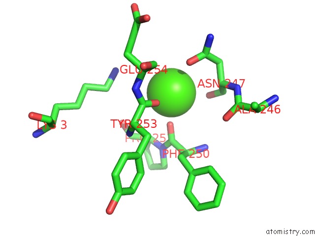
Mono view
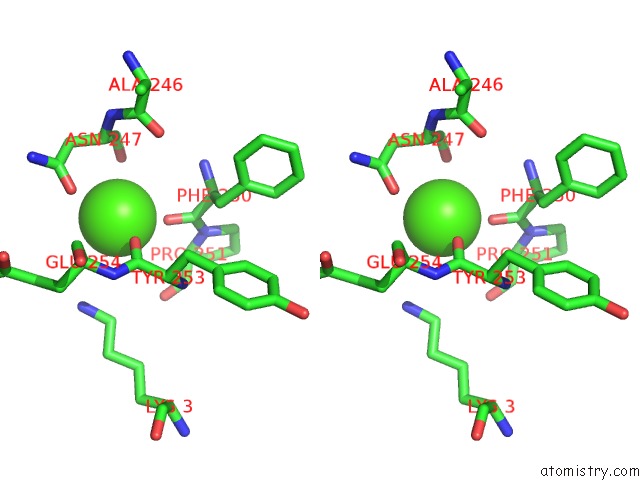
Stereo pair view
Mono view
Stereo pair view
A full contact list of Calcium with other atoms in the Ca binding
site number 1 of The Crystal Structure of A Protein in the Glycosyl Hydrolase Family 38 From Enterococcus Faecalis to 2.55A within 5.0Å range:
|
Reference:
A.J.Stein,
T.A.Binkowski,
A.Weger,
M.Borovilos,
S.Moy,
A.Joachimiak.
The Crystal Structure of A Protein in the Glycosyl Hydrolase Family 38 From Enterococcus Faecalis to 2.55A To Be Published.
Page generated: Tue Jul 8 14:25:58 2025
Last articles
Mg in 5D91Mg in 5D8G
Mg in 5D7R
Mg in 5D87
Mg in 5D5L
Mg in 5D86
Mg in 5D7D
Mg in 5D84
Mg in 5D7C
Mg in 5D7N