Calcium »
PDB 3m0c-3mip »
3mdo »
Calcium in PDB 3mdo: Crystal Structure of A Putative Phosphoribosylformylglycinamidine Cyclo-Ligase (BDI_2101) From Parabacteroides Distasonis Atcc 8503 at 1.91 A Resolution
Protein crystallography data
The structure of Crystal Structure of A Putative Phosphoribosylformylglycinamidine Cyclo-Ligase (BDI_2101) From Parabacteroides Distasonis Atcc 8503 at 1.91 A Resolution, PDB code: 3mdo
was solved by
Joint Center For Structural Genomics (Jcsg),
with X-Ray Crystallography technique. A brief refinement statistics is given in the table below:
| Resolution Low / High (Å) | 29.62 / 1.91 |
| Space group | P 41 21 2 |
| Cell size a, b, c (Å), α, β, γ (°) | 99.945, 99.945, 163.020, 90.00, 90.00, 90.00 |
| R / Rfree (%) | 15.4 / 19 |
Other elements in 3mdo:
The structure of Crystal Structure of A Putative Phosphoribosylformylglycinamidine Cyclo-Ligase (BDI_2101) From Parabacteroides Distasonis Atcc 8503 at 1.91 A Resolution also contains other interesting chemical elements:
| Arsenic | (As) | 1 atom |
| Chlorine | (Cl) | 4 atoms |
Calcium Binding Sites:
The binding sites of Calcium atom in the Crystal Structure of A Putative Phosphoribosylformylglycinamidine Cyclo-Ligase (BDI_2101) From Parabacteroides Distasonis Atcc 8503 at 1.91 A Resolution
(pdb code 3mdo). This binding sites where shown within
5.0 Angstroms radius around Calcium atom.
In total 7 binding sites of Calcium where determined in the Crystal Structure of A Putative Phosphoribosylformylglycinamidine Cyclo-Ligase (BDI_2101) From Parabacteroides Distasonis Atcc 8503 at 1.91 A Resolution, PDB code: 3mdo:
Jump to Calcium binding site number: 1; 2; 3; 4; 5; 6; 7;
In total 7 binding sites of Calcium where determined in the Crystal Structure of A Putative Phosphoribosylformylglycinamidine Cyclo-Ligase (BDI_2101) From Parabacteroides Distasonis Atcc 8503 at 1.91 A Resolution, PDB code: 3mdo:
Jump to Calcium binding site number: 1; 2; 3; 4; 5; 6; 7;
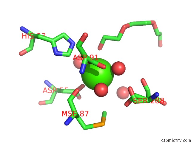
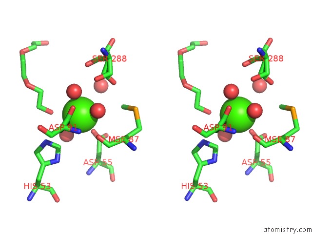
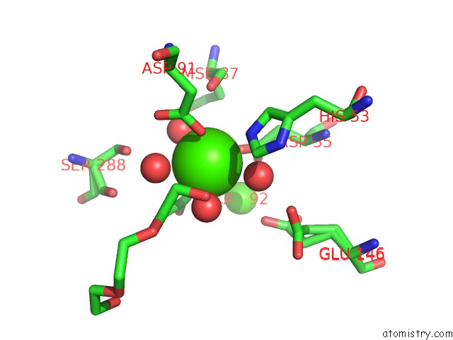
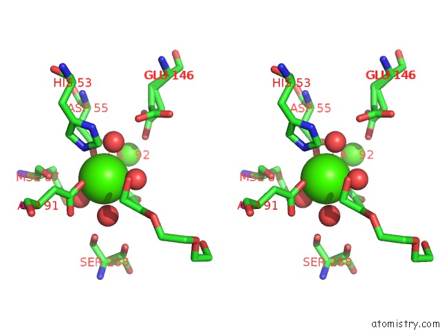
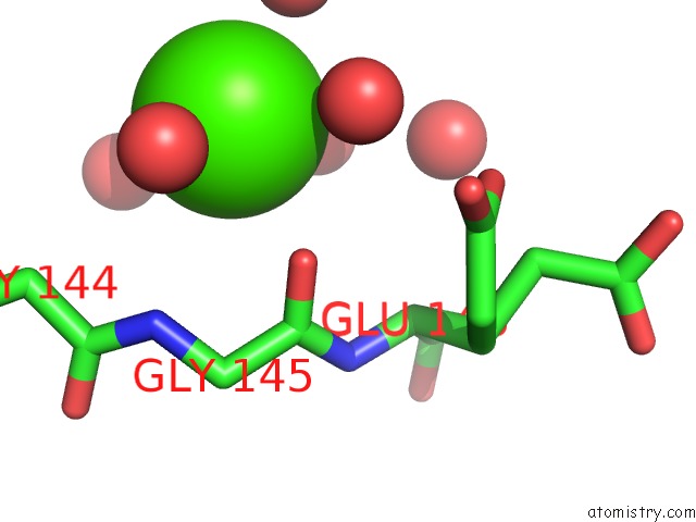
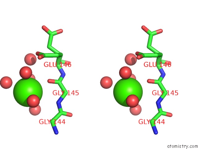
Calcium binding site 1 out of 7 in 3mdo
Go back to
Calcium binding site 1 out
of 7 in the Crystal Structure of A Putative Phosphoribosylformylglycinamidine Cyclo-Ligase (BDI_2101) From Parabacteroides Distasonis Atcc 8503 at 1.91 A Resolution
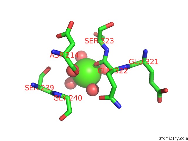
Mono view
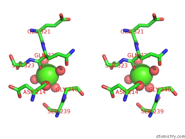
Stereo pair view
Mono view
Stereo pair view
A full contact list of Calcium with other atoms in the Ca binding
site number 1 of Crystal Structure of A Putative Phosphoribosylformylglycinamidine Cyclo-Ligase (BDI_2101) From Parabacteroides Distasonis Atcc 8503 at 1.91 A Resolution within 5.0Å range:
|
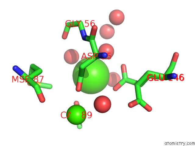
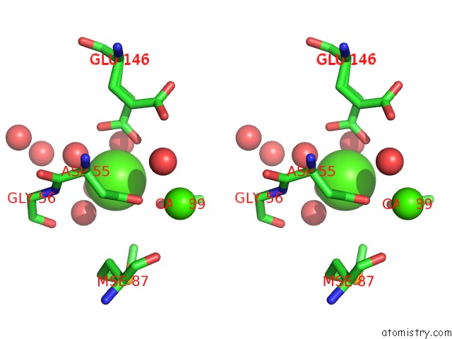
Calcium binding site 2 out of 7 in 3mdo
Go back to
Calcium binding site 2 out
of 7 in the Crystal Structure of A Putative Phosphoribosylformylglycinamidine Cyclo-Ligase (BDI_2101) From Parabacteroides Distasonis Atcc 8503 at 1.91 A Resolution
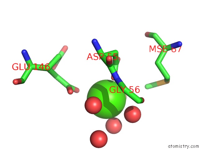
Mono view
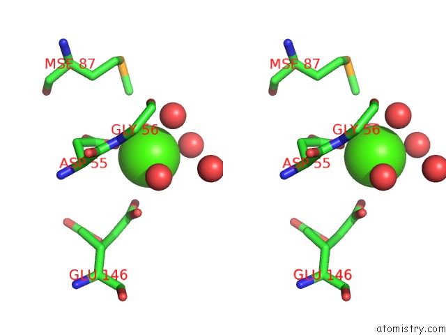
Stereo pair view
Mono view
Stereo pair view
A full contact list of Calcium with other atoms in the Ca binding
site number 2 of Crystal Structure of A Putative Phosphoribosylformylglycinamidine Cyclo-Ligase (BDI_2101) From Parabacteroides Distasonis Atcc 8503 at 1.91 A Resolution within 5.0Å range:
|
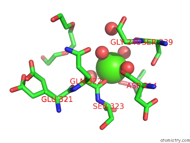
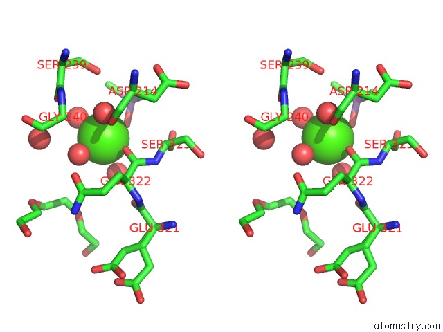
Calcium binding site 3 out of 7 in 3mdo
Go back to
Calcium binding site 3 out
of 7 in the Crystal Structure of A Putative Phosphoribosylformylglycinamidine Cyclo-Ligase (BDI_2101) From Parabacteroides Distasonis Atcc 8503 at 1.91 A Resolution
Mono view
Stereo pair view
Mono view
Stereo pair view
A full contact list of Calcium with other atoms in the Ca binding
site number 3 of Crystal Structure of A Putative Phosphoribosylformylglycinamidine Cyclo-Ligase (BDI_2101) From Parabacteroides Distasonis Atcc 8503 at 1.91 A Resolution within 5.0Å range:
|
Calcium binding site 4 out of 7 in 3mdo
Go back to
Calcium binding site 4 out
of 7 in the Crystal Structure of A Putative Phosphoribosylformylglycinamidine Cyclo-Ligase (BDI_2101) From Parabacteroides Distasonis Atcc 8503 at 1.91 A Resolution
Mono view
Stereo pair view
Mono view
Stereo pair view
A full contact list of Calcium with other atoms in the Ca binding
site number 4 of Crystal Structure of A Putative Phosphoribosylformylglycinamidine Cyclo-Ligase (BDI_2101) From Parabacteroides Distasonis Atcc 8503 at 1.91 A Resolution within 5.0Å range:
|
Calcium binding site 5 out of 7 in 3mdo
Go back to
Calcium binding site 5 out
of 7 in the Crystal Structure of A Putative Phosphoribosylformylglycinamidine Cyclo-Ligase (BDI_2101) From Parabacteroides Distasonis Atcc 8503 at 1.91 A Resolution
Mono view
Stereo pair view
Mono view
Stereo pair view
A full contact list of Calcium with other atoms in the Ca binding
site number 5 of Crystal Structure of A Putative Phosphoribosylformylglycinamidine Cyclo-Ligase (BDI_2101) From Parabacteroides Distasonis Atcc 8503 at 1.91 A Resolution within 5.0Å range:
|
Calcium binding site 6 out of 7 in 3mdo
Go back to
Calcium binding site 6 out
of 7 in the Crystal Structure of A Putative Phosphoribosylformylglycinamidine Cyclo-Ligase (BDI_2101) From Parabacteroides Distasonis Atcc 8503 at 1.91 A Resolution
Mono view
Stereo pair view
Mono view
Stereo pair view
A full contact list of Calcium with other atoms in the Ca binding
site number 6 of Crystal Structure of A Putative Phosphoribosylformylglycinamidine Cyclo-Ligase (BDI_2101) From Parabacteroides Distasonis Atcc 8503 at 1.91 A Resolution within 5.0Å range:
|
Calcium binding site 7 out of 7 in 3mdo
Go back to
Calcium binding site 7 out
of 7 in the Crystal Structure of A Putative Phosphoribosylformylglycinamidine Cyclo-Ligase (BDI_2101) From Parabacteroides Distasonis Atcc 8503 at 1.91 A Resolution
Mono view
Stereo pair view
Mono view
Stereo pair view
A full contact list of Calcium with other atoms in the Ca binding
site number 7 of Crystal Structure of A Putative Phosphoribosylformylglycinamidine Cyclo-Ligase (BDI_2101) From Parabacteroides Distasonis Atcc 8503 at 1.91 A Resolution within 5.0Å range:
|
Reference:
Joint Center For Structural Genomics (Jcsg),
Joint Center For Structural Genomics (Jcsg).
N/A N/A.
Page generated: Tue Jul 8 14:34:23 2025
Last articles
K in 5BTPK in 5BKE
K in 5BKK
K in 5BKJ
K in 5BJP
K in 5B2S
K in 5B2R
K in 5B2T
K in 5BJO
K in 5AW9