Calcium »
PDB 3p1o-3pdd »
3p7f »
Calcium in PDB 3p7f: Structure of the Human Langerin Carbohydrate Recognition Domain
Protein crystallography data
The structure of Structure of the Human Langerin Carbohydrate Recognition Domain, PDB code: 3p7f
was solved by
A.Skerra,
A.Schiefner,
with X-Ray Crystallography technique. A brief refinement statistics is given in the table below:
| Resolution Low / High (Å) | 28.20 / 2.50 |
| Space group | P 42 |
| Cell size a, b, c (Å), α, β, γ (°) | 79.760, 79.760, 90.560, 90.00, 90.00, 90.00 |
| R / Rfree (%) | 18.7 / 24.3 |
Calcium Binding Sites:
The binding sites of Calcium atom in the Structure of the Human Langerin Carbohydrate Recognition Domain
(pdb code 3p7f). This binding sites where shown within
5.0 Angstroms radius around Calcium atom.
In total 4 binding sites of Calcium where determined in the Structure of the Human Langerin Carbohydrate Recognition Domain, PDB code: 3p7f:
Jump to Calcium binding site number: 1; 2; 3; 4;
In total 4 binding sites of Calcium where determined in the Structure of the Human Langerin Carbohydrate Recognition Domain, PDB code: 3p7f:
Jump to Calcium binding site number: 1; 2; 3; 4;
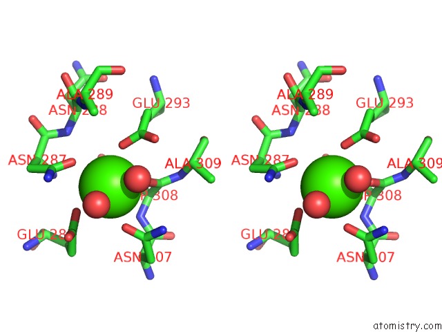
Calcium binding site 1 out of 4 in 3p7f
Go back to
Calcium binding site 1 out
of 4 in the Structure of the Human Langerin Carbohydrate Recognition Domain
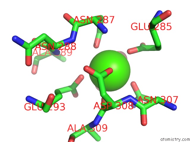
Mono view
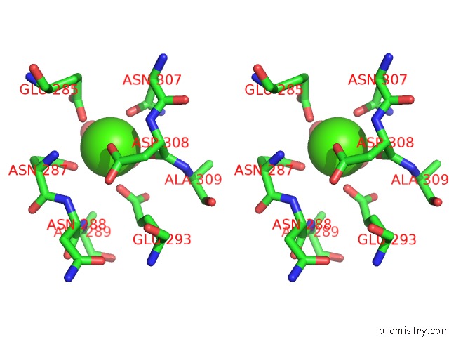
Stereo pair view
Mono view
Stereo pair view
A full contact list of Calcium with other atoms in the Ca binding
site number 1 of Structure of the Human Langerin Carbohydrate Recognition Domain within 5.0Å range:
|
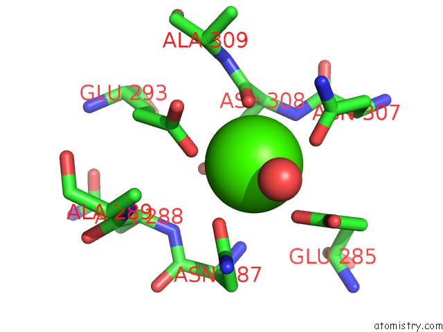
Calcium binding site 2 out of 4 in 3p7f
Go back to
Calcium binding site 2 out
of 4 in the Structure of the Human Langerin Carbohydrate Recognition Domain
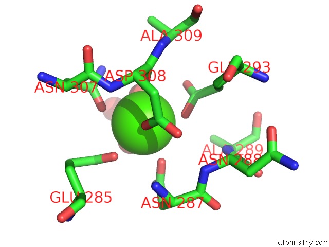
Mono view

Stereo pair view
Mono view
Stereo pair view
A full contact list of Calcium with other atoms in the Ca binding
site number 2 of Structure of the Human Langerin Carbohydrate Recognition Domain within 5.0Å range:
|
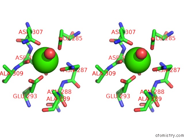
Calcium binding site 3 out of 4 in 3p7f
Go back to
Calcium binding site 3 out
of 4 in the Structure of the Human Langerin Carbohydrate Recognition Domain
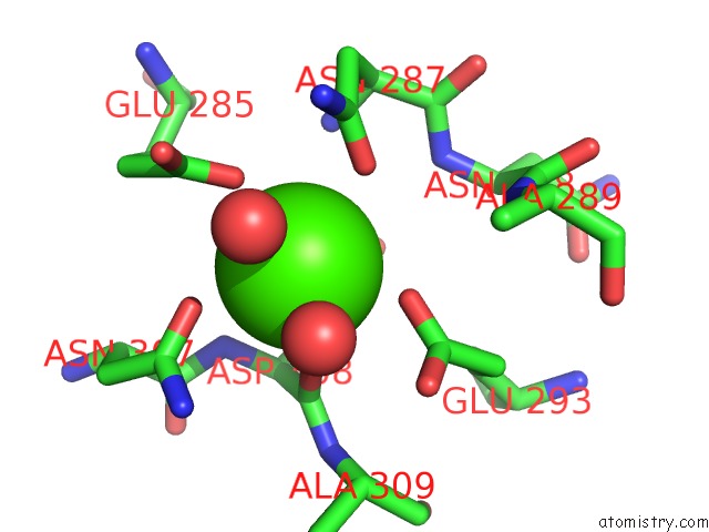
Mono view
Stereo pair view
Mono view
Stereo pair view
A full contact list of Calcium with other atoms in the Ca binding
site number 3 of Structure of the Human Langerin Carbohydrate Recognition Domain within 5.0Å range:
|
Calcium binding site 4 out of 4 in 3p7f
Go back to
Calcium binding site 4 out
of 4 in the Structure of the Human Langerin Carbohydrate Recognition Domain
Mono view
Stereo pair view
Mono view
Stereo pair view
A full contact list of Calcium with other atoms in the Ca binding
site number 4 of Structure of the Human Langerin Carbohydrate Recognition Domain within 5.0Å range:
|
Reference:
L.Chatwell,
A.Holla,
B.B.Kaufer,
A.Skerra.
The Carbohydrate Recognition Domain of Langerin Reveals High Structural Similarity with the One of Dc-Sign But An Additional, Calcium-Independent Sugar-Binding Site. Mol.Immunol. V. 45 1981 2008.
ISSN: ISSN 0161-5890
PubMed: 18061677
DOI: 10.1016/J.MOLIMM.2007.10.030
Page generated: Tue Jul 8 15:20:14 2025
ISSN: ISSN 0161-5890
PubMed: 18061677
DOI: 10.1016/J.MOLIMM.2007.10.030
Last articles
Mg in 7DU2Mg in 7DSP
Mg in 7DSJ
Mg in 7DSI
Mg in 7DRP
Mg in 7DSH
Mg in 7DSA
Mg in 7DRX
Mg in 7DRM
Mg in 7DPT