Calcium »
PDB 3pe0-3pty »
3pog »
Calcium in PDB 3pog: Crystal Structure of the Masp-1 CUB2 Domain Bound to CA2+
Protein crystallography data
The structure of Crystal Structure of the Masp-1 CUB2 Domain Bound to CA2+, PDB code: 3pog
was solved by
A.R.Gingras,
P.C.E.Moody,
R.Wallis,
with X-Ray Crystallography technique. A brief refinement statistics is given in the table below:
| Resolution Low / High (Å) | 38.98 / 2.75 |
| Space group | C 1 2 1 |
| Cell size a, b, c (Å), α, β, γ (°) | 111.680, 64.690, 52.420, 90.00, 92.33, 90.00 |
| R / Rfree (%) | 23.3 / 28.6 |
Calcium Binding Sites:
The binding sites of Calcium atom in the Crystal Structure of the Masp-1 CUB2 Domain Bound to CA2+
(pdb code 3pog). This binding sites where shown within
5.0 Angstroms radius around Calcium atom.
In total 3 binding sites of Calcium where determined in the Crystal Structure of the Masp-1 CUB2 Domain Bound to CA2+, PDB code: 3pog:
Jump to Calcium binding site number: 1; 2; 3;
In total 3 binding sites of Calcium where determined in the Crystal Structure of the Masp-1 CUB2 Domain Bound to CA2+, PDB code: 3pog:
Jump to Calcium binding site number: 1; 2; 3;
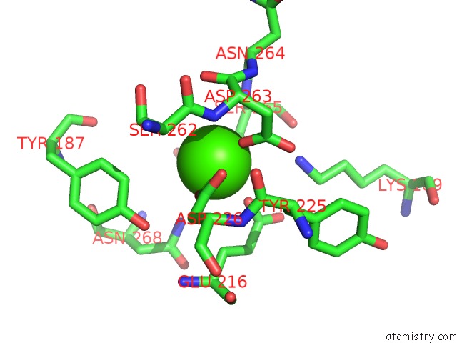
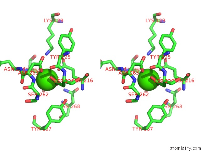
Calcium binding site 1 out of 3 in 3pog
Go back to
Calcium binding site 1 out
of 3 in the Crystal Structure of the Masp-1 CUB2 Domain Bound to CA2+
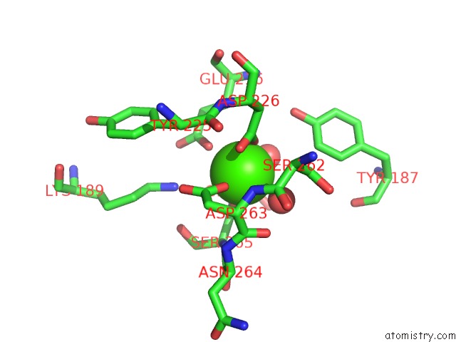
Mono view
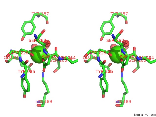
Stereo pair view
Mono view
Stereo pair view
A full contact list of Calcium with other atoms in the Ca binding
site number 1 of Crystal Structure of the Masp-1 CUB2 Domain Bound to CA2+ within 5.0Å range:
|
Calcium binding site 2 out of 3 in 3pog
Go back to
Calcium binding site 2 out
of 3 in the Crystal Structure of the Masp-1 CUB2 Domain Bound to CA2+
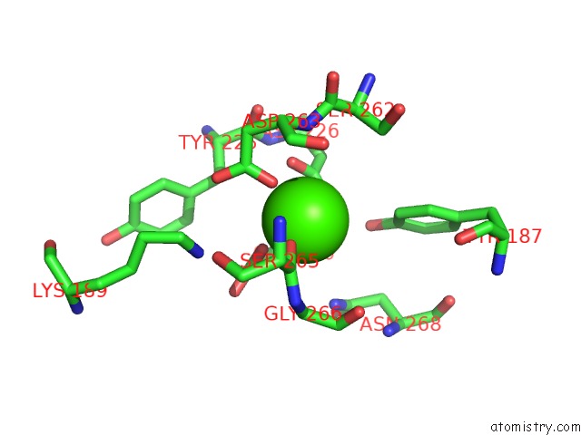
Mono view
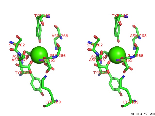
Stereo pair view
Mono view
Stereo pair view
A full contact list of Calcium with other atoms in the Ca binding
site number 2 of Crystal Structure of the Masp-1 CUB2 Domain Bound to CA2+ within 5.0Å range:
|
Calcium binding site 3 out of 3 in 3pog
Go back to
Calcium binding site 3 out
of 3 in the Crystal Structure of the Masp-1 CUB2 Domain Bound to CA2+
Mono view
Stereo pair view
Mono view
Stereo pair view
A full contact list of Calcium with other atoms in the Ca binding
site number 3 of Crystal Structure of the Masp-1 CUB2 Domain Bound to CA2+ within 5.0Å range:
|
Reference:
A.R.Gingras,
U.V.Girija,
A.H.Keeble,
R.Panchal,
D.A.Mitchell,
P.C.Moody,
R.Wallis.
Structural Basis of Mannan-Binding Lectin Recognition By Its Associated Serine Protease Masp-1: Implications For Complement Activation. Structure V. 19 1635 2011.
ISSN: ISSN 0969-2126
PubMed: 22078562
DOI: 10.1016/J.STR.2011.08.014
Page generated: Tue Jul 8 15:32:08 2025
ISSN: ISSN 0969-2126
PubMed: 22078562
DOI: 10.1016/J.STR.2011.08.014
Last articles
I in 8CEVI in 8C7R
I in 8C4W
I in 8C3L
I in 8BIR
I in 8C2G
I in 8BW9
I in 8BVX
I in 8ATE
I in 8AIL