Calcium »
PDB 3q4y-3qgo »
3qe2 »
Calcium in PDB 3qe2: Crystal Structure of Human Nadph-Cytochrome P450 Reductase
Enzymatic activity of Crystal Structure of Human Nadph-Cytochrome P450 Reductase
All present enzymatic activity of Crystal Structure of Human Nadph-Cytochrome P450 Reductase:
1.6.2.4;
1.6.2.4;
Protein crystallography data
The structure of Crystal Structure of Human Nadph-Cytochrome P450 Reductase, PDB code: 3qe2
was solved by
C.Xia,
C.Marohnic,
S.P.Panda,
B.S.Masters,
J.-J.P.Kim,
with X-Ray Crystallography technique. A brief refinement statistics is given in the table below:
| Resolution Low / High (Å) | 45.10 / 1.75 |
| Space group | P 21 21 21 |
| Cell size a, b, c (Å), α, β, γ (°) | 70.145, 117.782, 156.260, 90.00, 90.00, 90.00 |
| R / Rfree (%) | 21.1 / 23.9 |
Calcium Binding Sites:
The binding sites of Calcium atom in the Crystal Structure of Human Nadph-Cytochrome P450 Reductase
(pdb code 3qe2). This binding sites where shown within
5.0 Angstroms radius around Calcium atom.
In total only one binding site of Calcium was determined in the Crystal Structure of Human Nadph-Cytochrome P450 Reductase, PDB code: 3qe2:
In total only one binding site of Calcium was determined in the Crystal Structure of Human Nadph-Cytochrome P450 Reductase, PDB code: 3qe2:
Calcium binding site 1 out of 1 in 3qe2
Go back to
Calcium binding site 1 out
of 1 in the Crystal Structure of Human Nadph-Cytochrome P450 Reductase
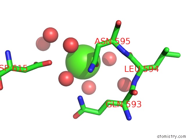
Mono view
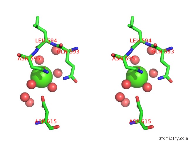
Stereo pair view
Mono view
Stereo pair view
A full contact list of Calcium with other atoms in the Ca binding
site number 1 of Crystal Structure of Human Nadph-Cytochrome P450 Reductase within 5.0Å range:
|
Reference:
C.Xia,
S.P.Panda,
C.C.Marohnic,
P.Martasek,
B.S.Masters,
J.J.Kim.
Structural Basis For Human Nadph-Cytochrome P450 Oxidoreductase Deficiency. Proc.Natl.Acad.Sci.Usa V. 108 13486 2011.
ISSN: ISSN 0027-8424
PubMed: 21808038
DOI: 10.1073/PNAS.1106632108
Page generated: Tue Jul 8 15:54:26 2025
ISSN: ISSN 0027-8424
PubMed: 21808038
DOI: 10.1073/PNAS.1106632108
Last articles
K in 4X3ZK in 4X3M
K in 4X16
K in 4X17
K in 4X3K
K in 4X12
K in 4X14
K in 4X15
K in 4X13
K in 4X11