Calcium »
PDB 3qgv-3r3t »
3qhq »
Calcium in PDB 3qhq: Structure of Crispr-Associated Protein CSN2
Enzymatic activity of Structure of Crispr-Associated Protein CSN2
All present enzymatic activity of Structure of Crispr-Associated Protein CSN2:
2.7.7.6;
2.7.7.6;
Protein crystallography data
The structure of Structure of Crispr-Associated Protein CSN2, PDB code: 3qhq
was solved by
P.Ellinger,
Z.Arslan,
R.Wurm,
B.Tschapek,
K.Pfeffer,
R.Wagner,
L.Schmitt,
U.Pul,
S.H.Smits,
with X-Ray Crystallography technique. A brief refinement statistics is given in the table below:
| Resolution Low / High (Å) | 19.57 / 2.00 |
| Space group | C 1 2 1 |
| Cell size a, b, c (Å), α, β, γ (°) | 75.300, 83.300, 110.400, 90.00, 109.40, 90.00 |
| R / Rfree (%) | 20.4 / 22.8 |
Calcium Binding Sites:
The binding sites of Calcium atom in the Structure of Crispr-Associated Protein CSN2
(pdb code 3qhq). This binding sites where shown within
5.0 Angstroms radius around Calcium atom.
In total 5 binding sites of Calcium where determined in the Structure of Crispr-Associated Protein CSN2, PDB code: 3qhq:
Jump to Calcium binding site number: 1; 2; 3; 4; 5;
In total 5 binding sites of Calcium where determined in the Structure of Crispr-Associated Protein CSN2, PDB code: 3qhq:
Jump to Calcium binding site number: 1; 2; 3; 4; 5;
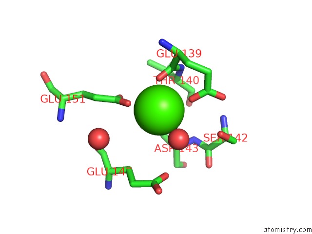
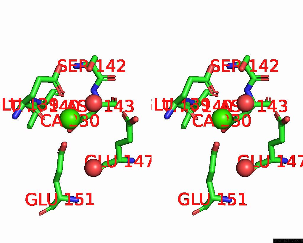
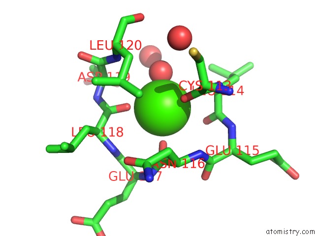
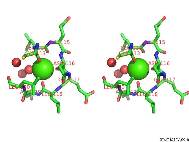
Calcium binding site 1 out of 5 in 3qhq
Go back to
Calcium binding site 1 out
of 5 in the Structure of Crispr-Associated Protein CSN2
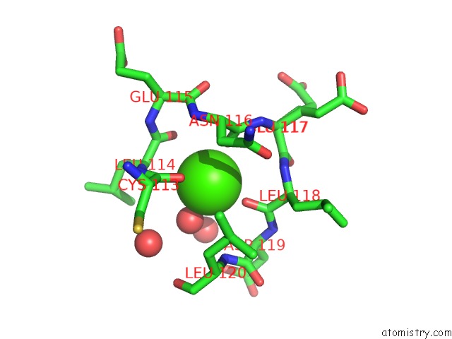
Mono view
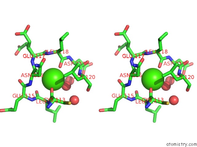
Stereo pair view
Mono view
Stereo pair view
A full contact list of Calcium with other atoms in the Ca binding
site number 1 of Structure of Crispr-Associated Protein CSN2 within 5.0Å range:
|
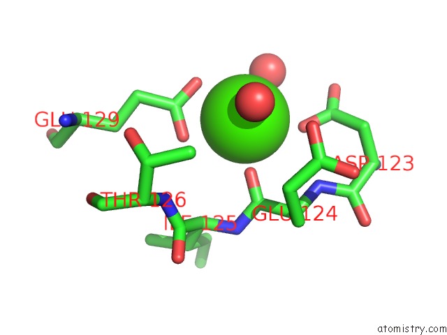
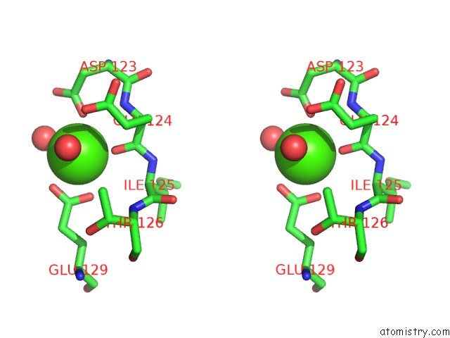
Calcium binding site 2 out of 5 in 3qhq
Go back to
Calcium binding site 2 out
of 5 in the Structure of Crispr-Associated Protein CSN2
Mono view
Stereo pair view
Mono view
Stereo pair view
A full contact list of Calcium with other atoms in the Ca binding
site number 2 of Structure of Crispr-Associated Protein CSN2 within 5.0Å range:
|
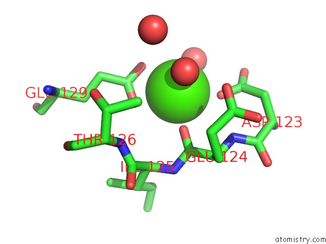
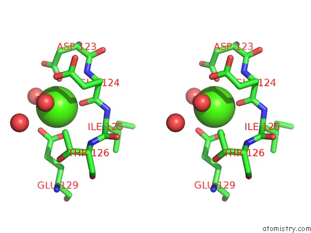
Calcium binding site 3 out of 5 in 3qhq
Go back to
Calcium binding site 3 out
of 5 in the Structure of Crispr-Associated Protein CSN2
Mono view
Stereo pair view
Mono view
Stereo pair view
A full contact list of Calcium with other atoms in the Ca binding
site number 3 of Structure of Crispr-Associated Protein CSN2 within 5.0Å range:
|
Calcium binding site 4 out of 5 in 3qhq
Go back to
Calcium binding site 4 out
of 5 in the Structure of Crispr-Associated Protein CSN2
Mono view
Stereo pair view
Mono view
Stereo pair view
A full contact list of Calcium with other atoms in the Ca binding
site number 4 of Structure of Crispr-Associated Protein CSN2 within 5.0Å range:
|
Calcium binding site 5 out of 5 in 3qhq
Go back to
Calcium binding site 5 out
of 5 in the Structure of Crispr-Associated Protein CSN2
Mono view
Stereo pair view
Mono view
Stereo pair view
A full contact list of Calcium with other atoms in the Ca binding
site number 5 of Structure of Crispr-Associated Protein CSN2 within 5.0Å range:
|
Reference:
P.Ellinger,
Z.Arslan,
R.Wurm,
B.Tschapek,
K.Pfeffer,
R.Wagner,
L.Schmitt,
U.Pul,
S.H.Smits.
Structure of Crispr-Associated Protein CSN2 To Be Published.
Page generated: Tue Jul 8 15:59:32 2025
Last articles
K in 6UB7K in 6U9P
K in 6U6C
K in 6U9L
K in 6U6D
K in 6U11
K in 6U98
K in 6TMX
K in 6U1K
K in 6U1J