Calcium »
PDB 3qgv-3r3t »
3qk1 »
Calcium in PDB 3qk1: Crystal Structure of Enterokinase-Like Trypsin Variant
Enzymatic activity of Crystal Structure of Enterokinase-Like Trypsin Variant
All present enzymatic activity of Crystal Structure of Enterokinase-Like Trypsin Variant:
3.4.21.4;
3.4.21.4;
Protein crystallography data
The structure of Crystal Structure of Enterokinase-Like Trypsin Variant, PDB code: 3qk1
was solved by
M.Schoepfel,
A.Tziridis,
U.Arnold,
M.T.Stubbs,
with X-Ray Crystallography technique. A brief refinement statistics is given in the table below:
| Resolution Low / High (Å) | 31.19 / 2.08 |
| Space group | P 21 21 21 |
| Cell size a, b, c (Å), α, β, γ (°) | 60.710, 63.760, 69.690, 90.00, 90.00, 90.00 |
| R / Rfree (%) | 18.2 / 23.7 |
Calcium Binding Sites:
The binding sites of Calcium atom in the Crystal Structure of Enterokinase-Like Trypsin Variant
(pdb code 3qk1). This binding sites where shown within
5.0 Angstroms radius around Calcium atom.
In total only one binding site of Calcium was determined in the Crystal Structure of Enterokinase-Like Trypsin Variant, PDB code: 3qk1:
In total only one binding site of Calcium was determined in the Crystal Structure of Enterokinase-Like Trypsin Variant, PDB code: 3qk1:
Calcium binding site 1 out of 1 in 3qk1
Go back to
Calcium binding site 1 out
of 1 in the Crystal Structure of Enterokinase-Like Trypsin Variant

Mono view
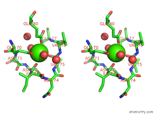
Stereo pair view
Mono view
Stereo pair view
A full contact list of Calcium with other atoms in the Ca binding
site number 1 of Crystal Structure of Enterokinase-Like Trypsin Variant within 5.0Å range:
|
Reference:
M.Schopfel,
A.Tziridis,
U.Arnold,
M.T.Stubbs.
Towards A Restriction Proteinase: Construction of A Self-Activating Enzyme. Chembiochem V. 12 1523 2011.
ISSN: ISSN 1439-4227
PubMed: 21387509
DOI: 10.1002/CBIC.201000787
Page generated: Tue Jul 8 16:00:31 2025
ISSN: ISSN 1439-4227
PubMed: 21387509
DOI: 10.1002/CBIC.201000787
Last articles
Fe in 2YXOFe in 2YRS
Fe in 2YXC
Fe in 2YNM
Fe in 2YVJ
Fe in 2YP1
Fe in 2YU2
Fe in 2YU1
Fe in 2YQB
Fe in 2YOO