Calcium »
PDB 3qgv-3r3t »
3qol »
Calcium in PDB 3qol: Crystal Structure of Staphylococcal Nuclease Variant D+Phs/V23E at pH 6 Determined at 100 K
Enzymatic activity of Crystal Structure of Staphylococcal Nuclease Variant D+Phs/V23E at pH 6 Determined at 100 K
All present enzymatic activity of Crystal Structure of Staphylococcal Nuclease Variant D+Phs/V23E at pH 6 Determined at 100 K:
3.1.31.1;
3.1.31.1;
Protein crystallography data
The structure of Crystal Structure of Staphylococcal Nuclease Variant D+Phs/V23E at pH 6 Determined at 100 K, PDB code: 3qol
was solved by
A.Robinson,
J.L.Schlessman,
E.B.Garcia-Moreno,
with X-Ray Crystallography technique. A brief refinement statistics is given in the table below:
| Resolution Low / High (Å) | 37.51 / 1.90 |
| Space group | P 1 21 1 |
| Cell size a, b, c (Å), α, β, γ (°) | 31.033, 60.453, 37.626, 90.00, 94.44, 90.00 |
| R / Rfree (%) | 18.7 / 22.7 |
Calcium Binding Sites:
The binding sites of Calcium atom in the Crystal Structure of Staphylococcal Nuclease Variant D+Phs/V23E at pH 6 Determined at 100 K
(pdb code 3qol). This binding sites where shown within
5.0 Angstroms radius around Calcium atom.
In total only one binding site of Calcium was determined in the Crystal Structure of Staphylococcal Nuclease Variant D+Phs/V23E at pH 6 Determined at 100 K, PDB code: 3qol:
In total only one binding site of Calcium was determined in the Crystal Structure of Staphylococcal Nuclease Variant D+Phs/V23E at pH 6 Determined at 100 K, PDB code: 3qol:
Calcium binding site 1 out of 1 in 3qol
Go back to
Calcium binding site 1 out
of 1 in the Crystal Structure of Staphylococcal Nuclease Variant D+Phs/V23E at pH 6 Determined at 100 K
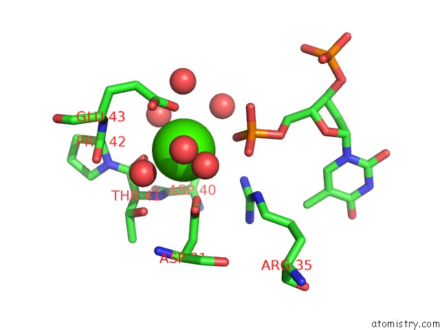
Mono view
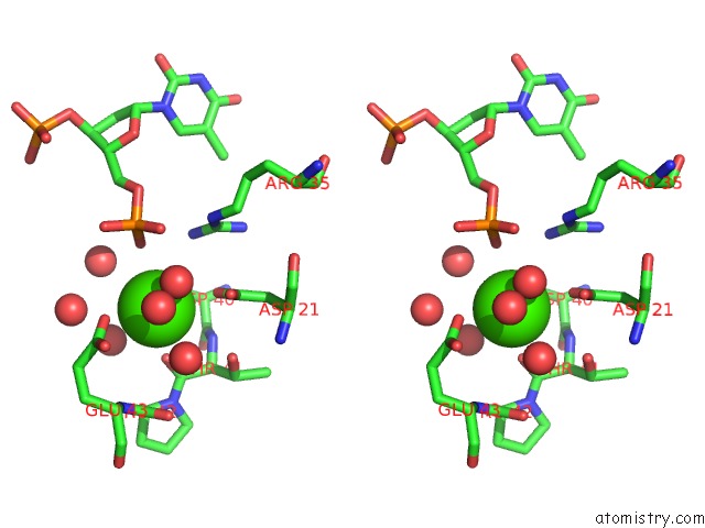
Stereo pair view
Mono view
Stereo pair view
A full contact list of Calcium with other atoms in the Ca binding
site number 1 of Crystal Structure of Staphylococcal Nuclease Variant D+Phs/V23E at pH 6 Determined at 100 K within 5.0Å range:
|
Reference:
A.C.Robinson,
C.A.Castaneda,
J.L.Schlessman,
B.Garcia-Moreno E.
Structural and Thermodynamic Consequences of Burial of An Artificial Ion Pair in the Hydrophobic Interior of A Protein. Proc.Natl.Acad.Sci.Usa V. 111 11685 2014.
ISSN: ISSN 0027-8424
PubMed: 25074910
DOI: 10.1073/PNAS.1402900111
Page generated: Tue Jul 8 16:02:21 2025
ISSN: ISSN 0027-8424
PubMed: 25074910
DOI: 10.1073/PNAS.1402900111
Last articles
K in 4BZ8K in 4C6V
K in 4C1L
K in 4C6U
K in 4C13
K in 4BZ7
K in 4C0O
K in 4BZ6
K in 4BZ5
K in 4BGU