Calcium »
PDB 3qgv-3r3t »
3qr7 »
Calcium in PDB 3qr7: Crystal Structure of the C-Terminal Fragment of the Bacteriophage P2 Membrane-Piercing Protein Gpv
Protein crystallography data
The structure of Crystal Structure of the C-Terminal Fragment of the Bacteriophage P2 Membrane-Piercing Protein Gpv, PDB code: 3qr7
was solved by
C.Browning,
M.Shneider,
P.G.Leiman,
with X-Ray Crystallography technique. A brief refinement statistics is given in the table below:
| Resolution Low / High (Å) | 27.33 / 0.94 |
| Space group | H 3 2 |
| Cell size a, b, c (Å), α, β, γ (°) | 49.069, 49.069, 463.896, 90.00, 90.00, 120.00 |
| R / Rfree (%) | 10.2 / 11.2 |
Other elements in 3qr7:
The structure of Crystal Structure of the C-Terminal Fragment of the Bacteriophage P2 Membrane-Piercing Protein Gpv also contains other interesting chemical elements:
| Iron | (Fe) | 2 atoms |
| Chlorine | (Cl) | 2 atoms |
| Sodium | (Na) | 1 atom |
Calcium Binding Sites:
The binding sites of Calcium atom in the Crystal Structure of the C-Terminal Fragment of the Bacteriophage P2 Membrane-Piercing Protein Gpv
(pdb code 3qr7). This binding sites where shown within
5.0 Angstroms radius around Calcium atom.
In total 2 binding sites of Calcium where determined in the Crystal Structure of the C-Terminal Fragment of the Bacteriophage P2 Membrane-Piercing Protein Gpv, PDB code: 3qr7:
Jump to Calcium binding site number: 1; 2;
In total 2 binding sites of Calcium where determined in the Crystal Structure of the C-Terminal Fragment of the Bacteriophage P2 Membrane-Piercing Protein Gpv, PDB code: 3qr7:
Jump to Calcium binding site number: 1; 2;
Calcium binding site 1 out of 2 in 3qr7
Go back to
Calcium binding site 1 out
of 2 in the Crystal Structure of the C-Terminal Fragment of the Bacteriophage P2 Membrane-Piercing Protein Gpv
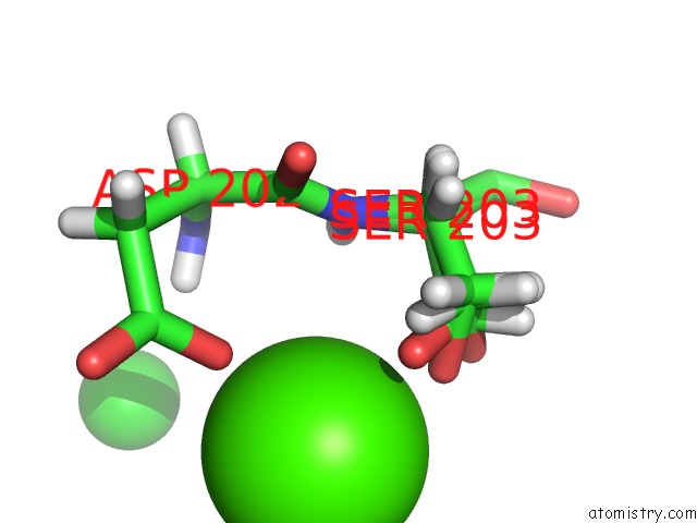
Mono view
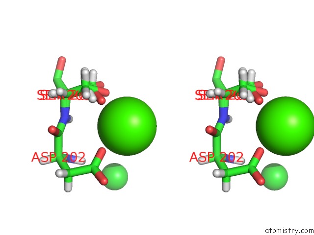
Stereo pair view
Mono view
Stereo pair view
A full contact list of Calcium with other atoms in the Ca binding
site number 1 of Crystal Structure of the C-Terminal Fragment of the Bacteriophage P2 Membrane-Piercing Protein Gpv within 5.0Å range:
|
Calcium binding site 2 out of 2 in 3qr7
Go back to
Calcium binding site 2 out
of 2 in the Crystal Structure of the C-Terminal Fragment of the Bacteriophage P2 Membrane-Piercing Protein Gpv
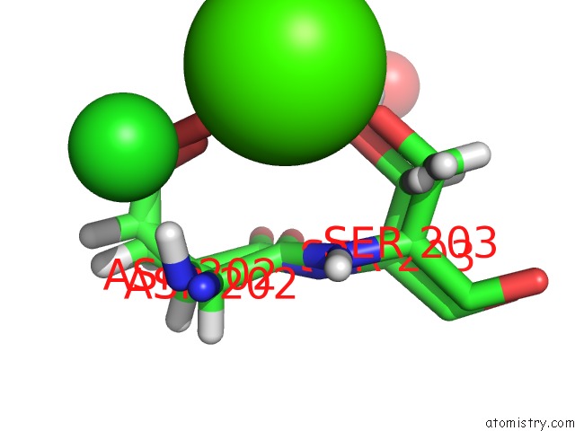
Mono view
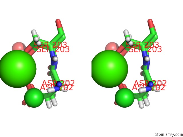
Stereo pair view
Mono view
Stereo pair view
A full contact list of Calcium with other atoms in the Ca binding
site number 2 of Crystal Structure of the C-Terminal Fragment of the Bacteriophage P2 Membrane-Piercing Protein Gpv within 5.0Å range:
|
Reference:
C.Browning,
M.M.Shneider,
V.D.Bowman,
D.Schwarzer,
P.G.Leiman.
Phage Pierces the Host Cell Membrane with the Iron-Loaded Spike. Structure V. 20 326 2012.
ISSN: ISSN 0969-2126
PubMed: 22325780
DOI: 10.1016/J.STR.2011.12.009
Page generated: Tue Jul 8 16:03:09 2025
ISSN: ISSN 0969-2126
PubMed: 22325780
DOI: 10.1016/J.STR.2011.12.009
Last articles
K in 4BZ9K in 4C6W
K in 4BZ8
K in 4C6V
K in 4C1L
K in 4C6U
K in 4C13
K in 4BZ7
K in 4C0O
K in 4BZ6