Calcium »
PDB 3r3v-3rk2 »
3rk2 »
Calcium in PDB 3rk2: Truncated Snare Complex
Protein crystallography data
The structure of Truncated Snare Complex, PDB code: 3rk2
was solved by
D.Kuemmel,
K.M.Reinisch,
with X-Ray Crystallography technique. A brief refinement statistics is given in the table below:
| Resolution Low / High (Å) | 39.50 / 2.20 |
| Space group | P 1 |
| Cell size a, b, c (Å), α, β, γ (°) | 27.627, 39.769, 102.275, 83.38, 89.94, 89.87 |
| R / Rfree (%) | 22.7 / 26.8 |
Calcium Binding Sites:
The binding sites of Calcium atom in the Truncated Snare Complex
(pdb code 3rk2). This binding sites where shown within
5.0 Angstroms radius around Calcium atom.
In total 4 binding sites of Calcium where determined in the Truncated Snare Complex, PDB code: 3rk2:
Jump to Calcium binding site number: 1; 2; 3; 4;
In total 4 binding sites of Calcium where determined in the Truncated Snare Complex, PDB code: 3rk2:
Jump to Calcium binding site number: 1; 2; 3; 4;
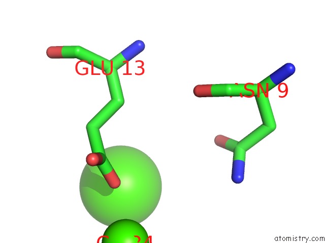
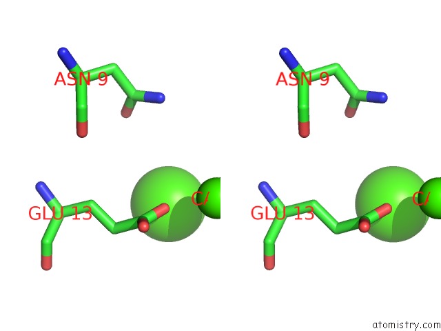
Calcium binding site 1 out of 4 in 3rk2
Go back to
Calcium binding site 1 out
of 4 in the Truncated Snare Complex
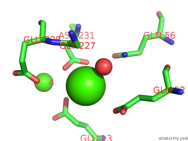
Mono view
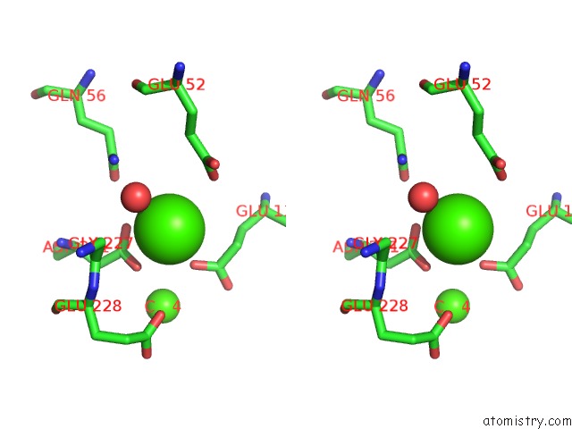
Stereo pair view
Mono view
Stereo pair view
A full contact list of Calcium with other atoms in the Ca binding
site number 1 of Truncated Snare Complex within 5.0Å range:
|
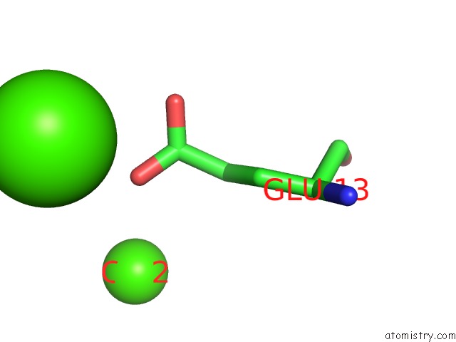
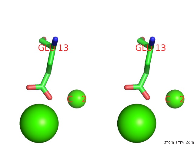
Calcium binding site 2 out of 4 in 3rk2
Go back to
Calcium binding site 2 out
of 4 in the Truncated Snare Complex
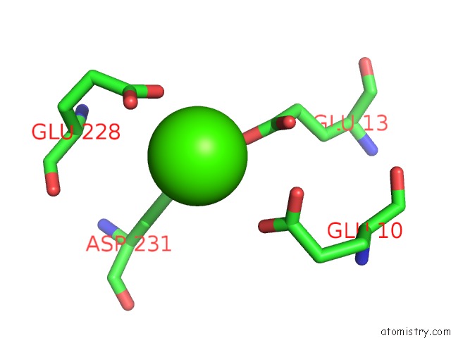
Mono view
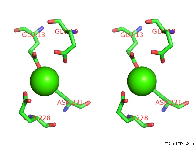
Stereo pair view
Mono view
Stereo pair view
A full contact list of Calcium with other atoms in the Ca binding
site number 2 of Truncated Snare Complex within 5.0Å range:
|
Calcium binding site 3 out of 4 in 3rk2
Go back to
Calcium binding site 3 out
of 4 in the Truncated Snare Complex
Mono view
Stereo pair view
Mono view
Stereo pair view
A full contact list of Calcium with other atoms in the Ca binding
site number 3 of Truncated Snare Complex within 5.0Å range:
|
Calcium binding site 4 out of 4 in 3rk2
Go back to
Calcium binding site 4 out
of 4 in the Truncated Snare Complex
Mono view
Stereo pair view
Mono view
Stereo pair view
A full contact list of Calcium with other atoms in the Ca binding
site number 4 of Truncated Snare Complex within 5.0Å range:
|
Reference:
D.Kummel,
S.S.Krishnakumar,
D.T.Radoff,
F.Li,
C.G.Giraudo,
F.Pincet,
J.E.Rothman,
K.M.Reinisch.
Complexin Cross-Links Prefusion Snares Into A Zigzag Array. Nat.Struct.Mol.Biol. V. 18 927 2011.
ISSN: ISSN 1545-9993
PubMed: 21785414
DOI: 10.1038/NSMB.2101
Page generated: Tue Jul 8 16:20:46 2025
ISSN: ISSN 1545-9993
PubMed: 21785414
DOI: 10.1038/NSMB.2101
Last articles
K in 8XW9K in 8XTS
K in 8XTR
K in 8XW7
K in 8XW6
K in 8XTQ
K in 8XMI
K in 8XTP
K in 8XMH
K in 8X9Q