Calcium »
PDB 3ti4-3tvc »
3tpq »
Calcium in PDB 3tpq: Crystal Structure of Wild-Type Mal Rpel Domain in Complex with Five G- Actins
Protein crystallography data
The structure of Crystal Structure of Wild-Type Mal Rpel Domain in Complex with Five G- Actins, PDB code: 3tpq
was solved by
H.Hirano,
Y.Matsuura,
with X-Ray Crystallography technique. A brief refinement statistics is given in the table below:
| Resolution Low / High (Å) | 68.70 / 3.45 |
| Space group | P 65 2 2 |
| Cell size a, b, c (Å), α, β, γ (°) | 180.737, 180.737, 382.279, 90.00, 90.00, 120.00 |
| R / Rfree (%) | 22.4 / 27.3 |
Calcium Binding Sites:
The binding sites of Calcium atom in the Crystal Structure of Wild-Type Mal Rpel Domain in Complex with Five G- Actins
(pdb code 3tpq). This binding sites where shown within
5.0 Angstroms radius around Calcium atom.
In total 5 binding sites of Calcium where determined in the Crystal Structure of Wild-Type Mal Rpel Domain in Complex with Five G- Actins, PDB code: 3tpq:
Jump to Calcium binding site number: 1; 2; 3; 4; 5;
In total 5 binding sites of Calcium where determined in the Crystal Structure of Wild-Type Mal Rpel Domain in Complex with Five G- Actins, PDB code: 3tpq:
Jump to Calcium binding site number: 1; 2; 3; 4; 5;
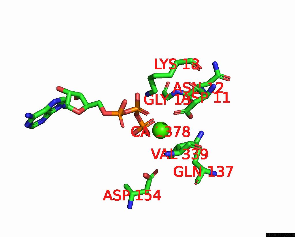
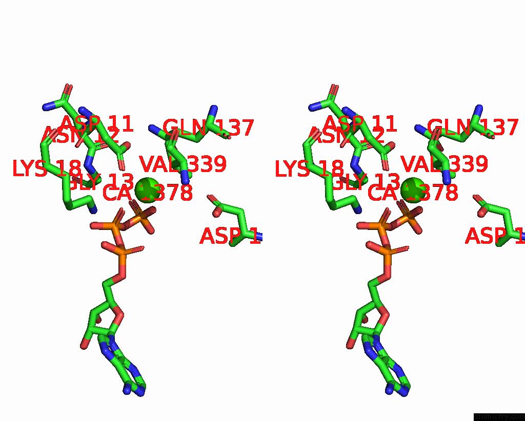
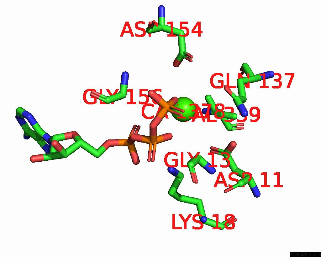
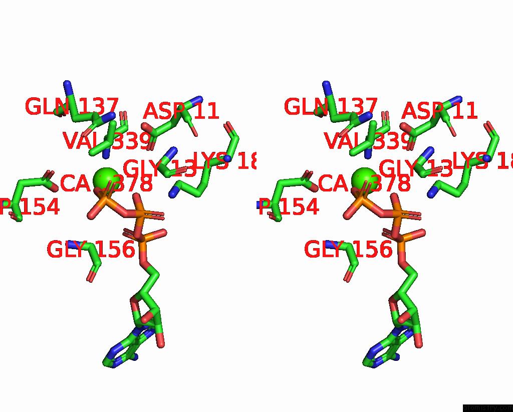
Calcium binding site 1 out of 5 in 3tpq
Go back to
Calcium binding site 1 out
of 5 in the Crystal Structure of Wild-Type Mal Rpel Domain in Complex with Five G- Actins
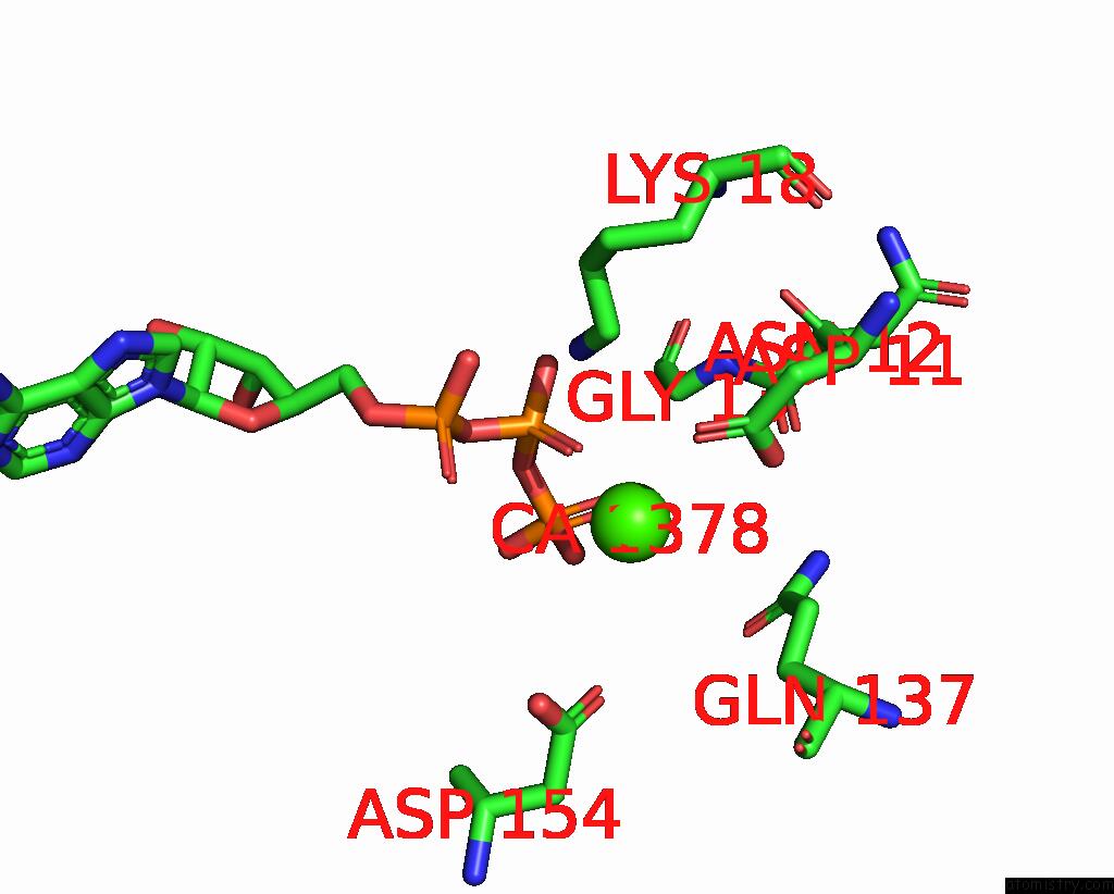
Mono view
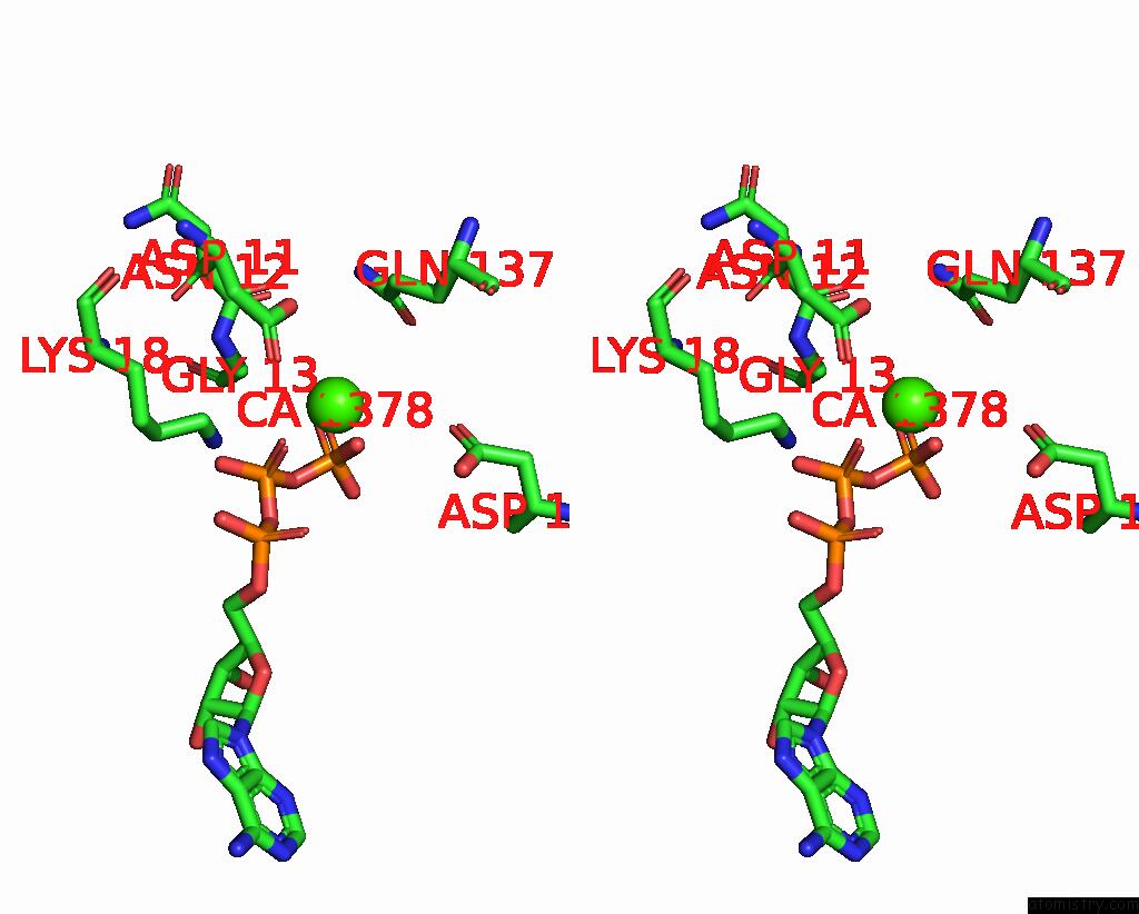
Stereo pair view
Mono view
Stereo pair view
A full contact list of Calcium with other atoms in the Ca binding
site number 1 of Crystal Structure of Wild-Type Mal Rpel Domain in Complex with Five G- Actins within 5.0Å range:
|
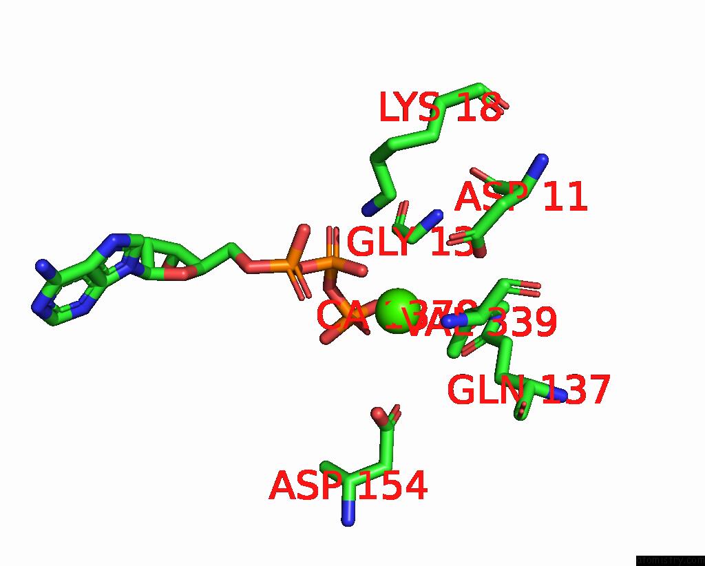
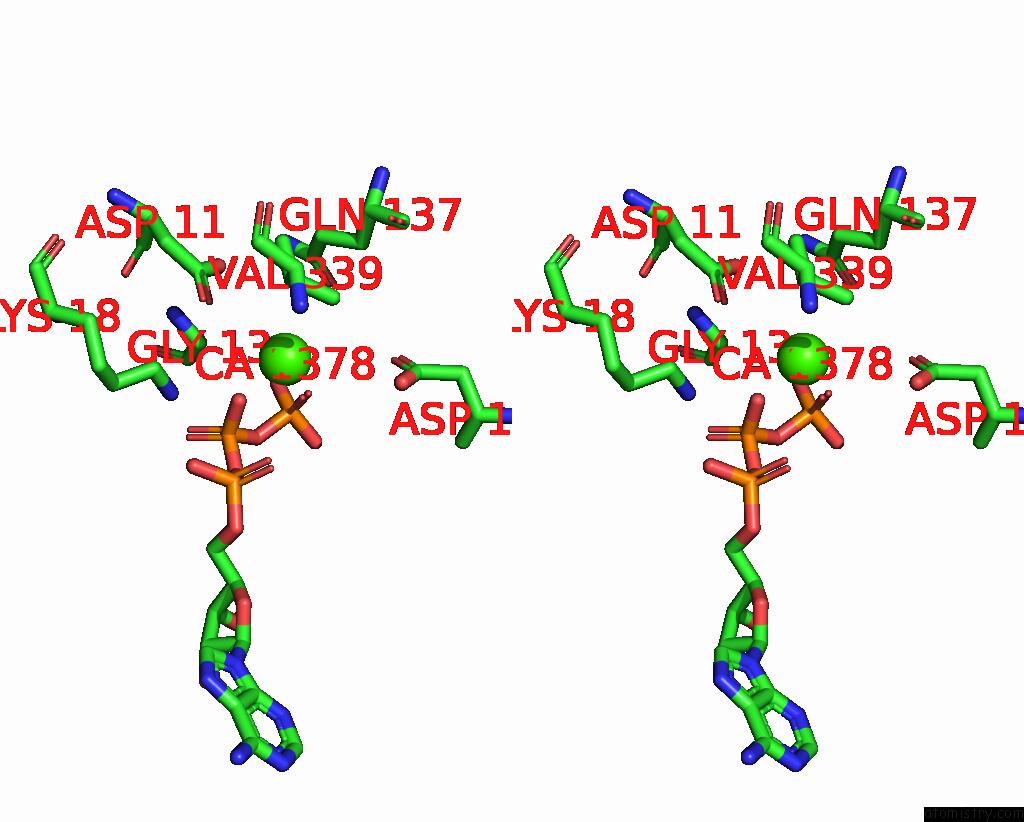
Calcium binding site 2 out of 5 in 3tpq
Go back to
Calcium binding site 2 out
of 5 in the Crystal Structure of Wild-Type Mal Rpel Domain in Complex with Five G- Actins
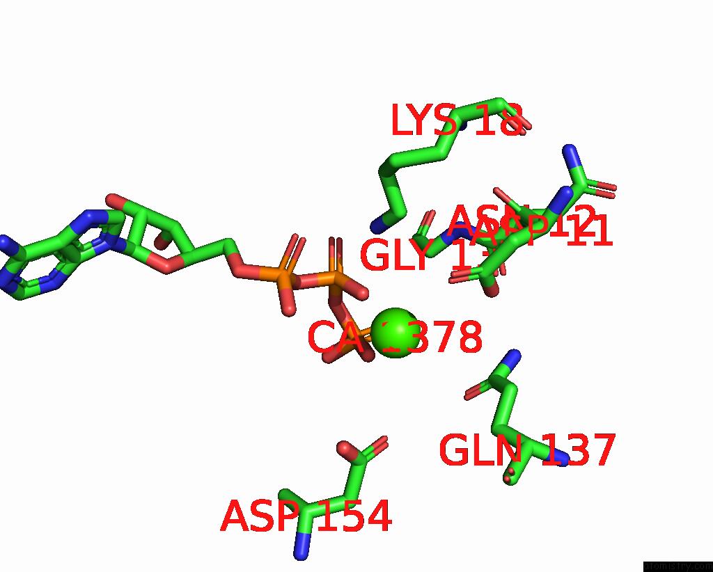
Mono view
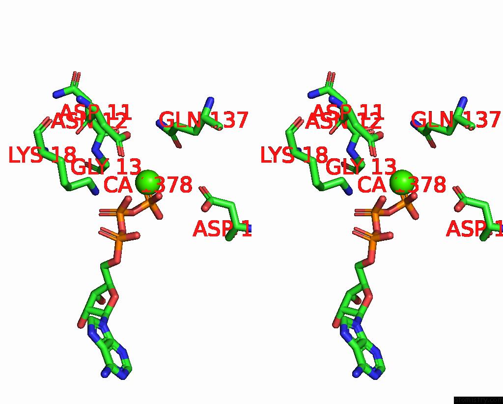
Stereo pair view
Mono view
Stereo pair view
A full contact list of Calcium with other atoms in the Ca binding
site number 2 of Crystal Structure of Wild-Type Mal Rpel Domain in Complex with Five G- Actins within 5.0Å range:
|
Calcium binding site 3 out of 5 in 3tpq
Go back to
Calcium binding site 3 out
of 5 in the Crystal Structure of Wild-Type Mal Rpel Domain in Complex with Five G- Actins
Mono view
Stereo pair view
Mono view
Stereo pair view
A full contact list of Calcium with other atoms in the Ca binding
site number 3 of Crystal Structure of Wild-Type Mal Rpel Domain in Complex with Five G- Actins within 5.0Å range:
|
Calcium binding site 4 out of 5 in 3tpq
Go back to
Calcium binding site 4 out
of 5 in the Crystal Structure of Wild-Type Mal Rpel Domain in Complex with Five G- Actins
Mono view
Stereo pair view
Mono view
Stereo pair view
A full contact list of Calcium with other atoms in the Ca binding
site number 4 of Crystal Structure of Wild-Type Mal Rpel Domain in Complex with Five G- Actins within 5.0Å range:
|
Calcium binding site 5 out of 5 in 3tpq
Go back to
Calcium binding site 5 out
of 5 in the Crystal Structure of Wild-Type Mal Rpel Domain in Complex with Five G- Actins
Mono view
Stereo pair view
Mono view
Stereo pair view
A full contact list of Calcium with other atoms in the Ca binding
site number 5 of Crystal Structure of Wild-Type Mal Rpel Domain in Complex with Five G- Actins within 5.0Å range:
|
Reference:
H.Hirano,
Y.Matsuura.
Sensing Actin Dynamics: Structural Basis For G-Actin-Sensitive Nuclear Import of Mal Biochem.Biophys.Res.Commun. V. 414 373 2011.
ISSN: ISSN 0006-291X
PubMed: 21964294
DOI: 10.1016/J.BBRC.2011.09.079
Page generated: Sat Jul 13 19:52:14 2024
ISSN: ISSN 0006-291X
PubMed: 21964294
DOI: 10.1016/J.BBRC.2011.09.079
Last articles
Zn in 9MJ5Zn in 9HNW
Zn in 9G0L
Zn in 9FNE
Zn in 9DZN
Zn in 9E0I
Zn in 9D32
Zn in 9DAK
Zn in 8ZXC
Zn in 8ZUF