Calcium »
PDB 3tz1-3uiq »
3u43 »
Calcium in PDB 3u43: Crystal Structure of the Colicin E2 Dnase-IM2 Complex
Protein crystallography data
The structure of Crystal Structure of the Colicin E2 Dnase-IM2 Complex, PDB code: 3u43
was solved by
J.A.Wojdyla,
C.Kleanthous,
with X-Ray Crystallography technique. A brief refinement statistics is given in the table below:
| Resolution Low / High (Å) | 48.81 / 1.72 |
| Space group | P 21 21 2 |
| Cell size a, b, c (Å), α, β, γ (°) | 121.814, 53.277, 32.785, 90.00, 90.00, 90.00 |
| R / Rfree (%) | 16.2 / 20.3 |
Other elements in 3u43:
The structure of Crystal Structure of the Colicin E2 Dnase-IM2 Complex also contains other interesting chemical elements:
| Zinc | (Zn) | 1 atom |
Calcium Binding Sites:
The binding sites of Calcium atom in the Crystal Structure of the Colicin E2 Dnase-IM2 Complex
(pdb code 3u43). This binding sites where shown within
5.0 Angstroms radius around Calcium atom.
In total only one binding site of Calcium was determined in the Crystal Structure of the Colicin E2 Dnase-IM2 Complex, PDB code: 3u43:
In total only one binding site of Calcium was determined in the Crystal Structure of the Colicin E2 Dnase-IM2 Complex, PDB code: 3u43:
Calcium binding site 1 out of 1 in 3u43
Go back to
Calcium binding site 1 out
of 1 in the Crystal Structure of the Colicin E2 Dnase-IM2 Complex
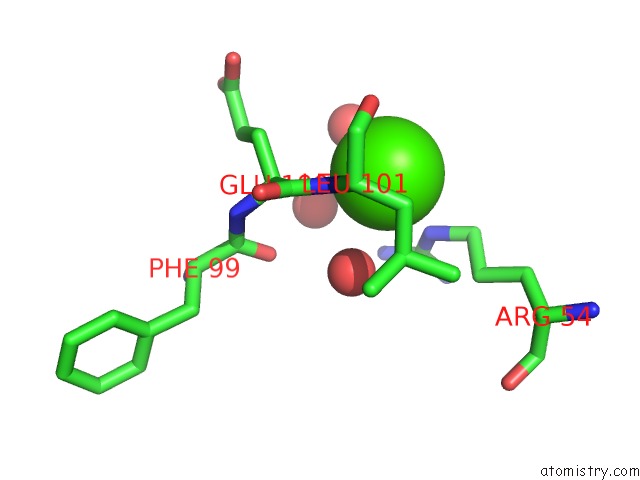
Mono view
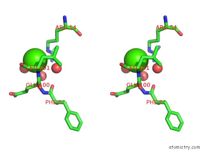
Stereo pair view
Mono view
Stereo pair view
A full contact list of Calcium with other atoms in the Ca binding
site number 1 of Crystal Structure of the Colicin E2 Dnase-IM2 Complex within 5.0Å range:
|
Reference:
J.A.Wojdyla,
S.J.Fleishman,
D.Baker,
C.Kleanthous.
Structure of the Ultra-High-Affinity Colicin E2 Dnase-IM2 Complex. J.Mol.Biol. V. 417 79 2012.
ISSN: ISSN 0022-2836
PubMed: 22306467
DOI: 10.1016/J.JMB.2012.01.019
Page generated: Tue Jul 8 17:09:28 2025
ISSN: ISSN 0022-2836
PubMed: 22306467
DOI: 10.1016/J.JMB.2012.01.019
Last articles
Fe in 7R2SFe in 7R0W
Fe in 7R2R
Fe in 7R2P
Fe in 7R2O
Fe in 7R1J
Fe in 7R1I
Fe in 7QWT
Fe in 7R1H
Fe in 7R0F