Calcium »
PDB 3tz1-3uiq »
3uba »
Calcium in PDB 3uba: Crystal Structure of the Complex of Bovine Lactoperoxidase with P- Hydroxycinnamic Acid at 2.6 A Resolution
Enzymatic activity of Crystal Structure of the Complex of Bovine Lactoperoxidase with P- Hydroxycinnamic Acid at 2.6 A Resolution
All present enzymatic activity of Crystal Structure of the Complex of Bovine Lactoperoxidase with P- Hydroxycinnamic Acid at 2.6 A Resolution:
1.11.1.7;
1.11.1.7;
Protein crystallography data
The structure of Crystal Structure of the Complex of Bovine Lactoperoxidase with P- Hydroxycinnamic Acid at 2.6 A Resolution, PDB code: 3uba
was solved by
N.Pandey,
A.K.Singh,
R.P.Singh,
M.Sinha,
P.Kaur,
S.Sharma,
T.P.Singh,
with X-Ray Crystallography technique. A brief refinement statistics is given in the table below:
| Resolution Low / High (Å) | 43.99 / 2.65 |
| Space group | P 1 21 1 |
| Cell size a, b, c (Å), α, β, γ (°) | 54.050, 80.230, 76.100, 90.00, 103.33, 90.00 |
| R / Rfree (%) | 21.9 / 23.8 |
Other elements in 3uba:
The structure of Crystal Structure of the Complex of Bovine Lactoperoxidase with P- Hydroxycinnamic Acid at 2.6 A Resolution also contains other interesting chemical elements:
| Iodine | (I) | 9 atoms |
| Iron | (Fe) | 1 atom |
Calcium Binding Sites:
The binding sites of Calcium atom in the Crystal Structure of the Complex of Bovine Lactoperoxidase with P- Hydroxycinnamic Acid at 2.6 A Resolution
(pdb code 3uba). This binding sites where shown within
5.0 Angstroms radius around Calcium atom.
In total only one binding site of Calcium was determined in the Crystal Structure of the Complex of Bovine Lactoperoxidase with P- Hydroxycinnamic Acid at 2.6 A Resolution, PDB code: 3uba:
In total only one binding site of Calcium was determined in the Crystal Structure of the Complex of Bovine Lactoperoxidase with P- Hydroxycinnamic Acid at 2.6 A Resolution, PDB code: 3uba:
Calcium binding site 1 out of 1 in 3uba
Go back to
Calcium binding site 1 out
of 1 in the Crystal Structure of the Complex of Bovine Lactoperoxidase with P- Hydroxycinnamic Acid at 2.6 A Resolution
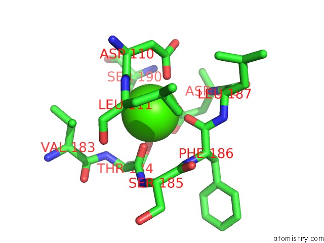
Mono view
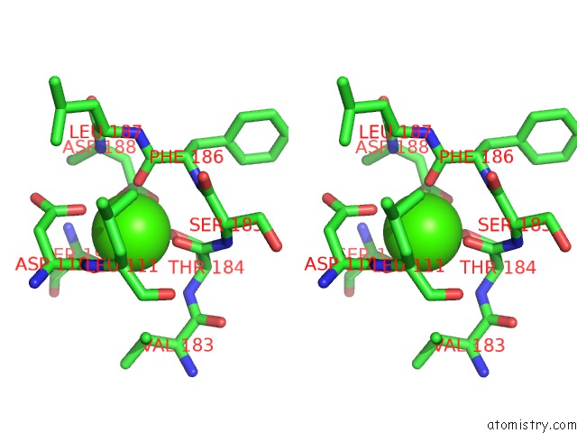
Stereo pair view
Mono view
Stereo pair view
A full contact list of Calcium with other atoms in the Ca binding
site number 1 of Crystal Structure of the Complex of Bovine Lactoperoxidase with P- Hydroxycinnamic Acid at 2.6 A Resolution within 5.0Å range:
|
Reference:
N.Pandey,
A.K.Singh,
R.P.Singh,
M.Sinha,
P.Kaur,
S.Sharma,
T.P.Singh.
Crystal Structure of the Complex of Bovine Lactoperoxidase with P-Hydroxycinnamic Acid at 2.6 A Resolution To Be Published.
Page generated: Tue Jul 8 17:13:11 2025
Last articles
Fe in 7R2SFe in 7R0W
Fe in 7R2R
Fe in 7R2P
Fe in 7R2O
Fe in 7R1J
Fe in 7R1I
Fe in 7QWT
Fe in 7R1H
Fe in 7R0F