Calcium »
PDB 3tz1-3uiq »
3uho »
Calcium in PDB 3uho: Crystal Structure of Glutamate Racemase From Campylobacter Jejuni Subsp. Jejuni
Enzymatic activity of Crystal Structure of Glutamate Racemase From Campylobacter Jejuni Subsp. Jejuni
All present enzymatic activity of Crystal Structure of Glutamate Racemase From Campylobacter Jejuni Subsp. Jejuni:
5.1.1.3;
5.1.1.3;
Protein crystallography data
The structure of Crystal Structure of Glutamate Racemase From Campylobacter Jejuni Subsp. Jejuni, PDB code: 3uho
was solved by
N.Maltseva,
R.Mulligan,
K.Kwon,
Y.Kim,
W.F.Anderson,
A.Joachimiak,
Centerfor Structural Genomics Of Infectious Diseases (Csgid),
with X-Ray Crystallography technique. A brief refinement statistics is given in the table below:
| Resolution Low / High (Å) | 34.60 / 2.20 |
| Space group | C 1 2 1 |
| Cell size a, b, c (Å), α, β, γ (°) | 139.308, 74.384, 50.144, 90.00, 96.53, 90.00 |
| R / Rfree (%) | 19 / 23.6 |
Other elements in 3uho:
The structure of Crystal Structure of Glutamate Racemase From Campylobacter Jejuni Subsp. Jejuni also contains other interesting chemical elements:
| Chlorine | (Cl) | 3 atoms |
Calcium Binding Sites:
The binding sites of Calcium atom in the Crystal Structure of Glutamate Racemase From Campylobacter Jejuni Subsp. Jejuni
(pdb code 3uho). This binding sites where shown within
5.0 Angstroms radius around Calcium atom.
In total 2 binding sites of Calcium where determined in the Crystal Structure of Glutamate Racemase From Campylobacter Jejuni Subsp. Jejuni, PDB code: 3uho:
Jump to Calcium binding site number: 1; 2;
In total 2 binding sites of Calcium where determined in the Crystal Structure of Glutamate Racemase From Campylobacter Jejuni Subsp. Jejuni, PDB code: 3uho:
Jump to Calcium binding site number: 1; 2;
Calcium binding site 1 out of 2 in 3uho
Go back to
Calcium binding site 1 out
of 2 in the Crystal Structure of Glutamate Racemase From Campylobacter Jejuni Subsp. Jejuni
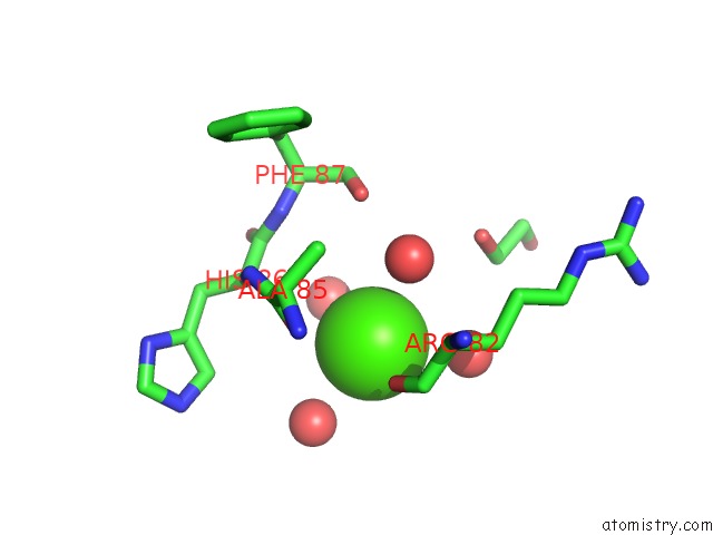
Mono view
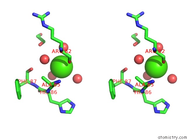
Stereo pair view
Mono view
Stereo pair view
A full contact list of Calcium with other atoms in the Ca binding
site number 1 of Crystal Structure of Glutamate Racemase From Campylobacter Jejuni Subsp. Jejuni within 5.0Å range:
|
Calcium binding site 2 out of 2 in 3uho
Go back to
Calcium binding site 2 out
of 2 in the Crystal Structure of Glutamate Racemase From Campylobacter Jejuni Subsp. Jejuni
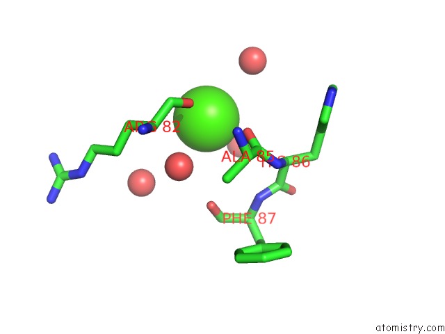
Mono view
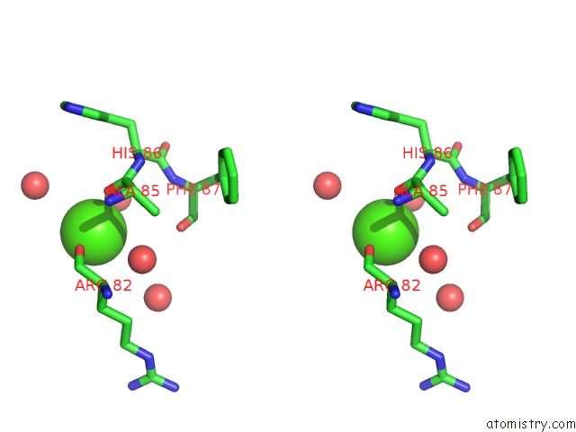
Stereo pair view
Mono view
Stereo pair view
A full contact list of Calcium with other atoms in the Ca binding
site number 2 of Crystal Structure of Glutamate Racemase From Campylobacter Jejuni Subsp. Jejuni within 5.0Å range:
|
Reference:
N.Maltseva,
R.Mulligan,
K.Kwon,
Y.Kim,
W.F.Anderson,
A.Joachimiak,
Csgid.
Crystal Structure of Glutamate Racemase From Campylobacter Jejuni Subsp. Jejuni To Be Published.
Page generated: Tue Jul 8 17:15:09 2025
Last articles
Mg in 6SU2Mg in 6STF
Mg in 6STG
Mg in 6ST5
Mg in 6SQZ
Mg in 6SR6
Mg in 6SQY
Mg in 6SQW
Mg in 6SQJ
Mg in 6SQ2