Calcium »
PDB 3v0a-3vl2 »
3v7f »
Calcium in PDB 3v7f: Crystal Structure of Streptococcus Pyogenes CSN2
Protein crystallography data
The structure of Crystal Structure of Streptococcus Pyogenes CSN2, PDB code: 3v7f
was solved by
E.Bae,
D.K.Jung,
Y.Koo,
with X-Ray Crystallography technique. A brief refinement statistics is given in the table below:
| Resolution Low / High (Å) | 49.68 / 2.90 |
| Space group | C 2 2 21 |
| Cell size a, b, c (Å), α, β, γ (°) | 59.060, 162.920, 149.040, 90.00, 90.00, 90.00 |
| R / Rfree (%) | 21.6 / 26.7 |
Calcium Binding Sites:
The binding sites of Calcium atom in the Crystal Structure of Streptococcus Pyogenes CSN2
(pdb code 3v7f). This binding sites where shown within
5.0 Angstroms radius around Calcium atom.
In total 3 binding sites of Calcium where determined in the Crystal Structure of Streptococcus Pyogenes CSN2, PDB code: 3v7f:
Jump to Calcium binding site number: 1; 2; 3;
In total 3 binding sites of Calcium where determined in the Crystal Structure of Streptococcus Pyogenes CSN2, PDB code: 3v7f:
Jump to Calcium binding site number: 1; 2; 3;
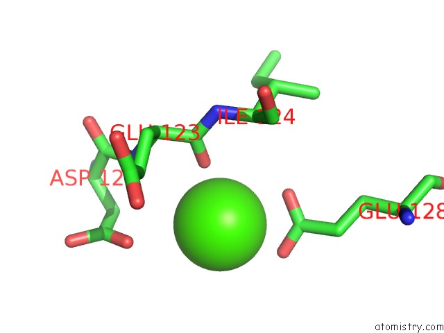
Calcium binding site 1 out of 3 in 3v7f
Go back to
Calcium binding site 1 out
of 3 in the Crystal Structure of Streptococcus Pyogenes CSN2

Mono view
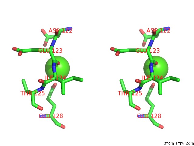
Stereo pair view
Mono view
Stereo pair view
A full contact list of Calcium with other atoms in the Ca binding
site number 1 of Crystal Structure of Streptococcus Pyogenes CSN2 within 5.0Å range:
|
Calcium binding site 2 out of 3 in 3v7f
Go back to
Calcium binding site 2 out
of 3 in the Crystal Structure of Streptococcus Pyogenes CSN2

Mono view
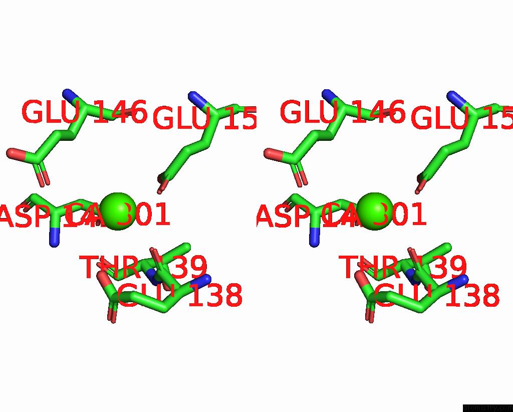
Stereo pair view
Mono view
Stereo pair view
A full contact list of Calcium with other atoms in the Ca binding
site number 2 of Crystal Structure of Streptococcus Pyogenes CSN2 within 5.0Å range:
|
Calcium binding site 3 out of 3 in 3v7f
Go back to
Calcium binding site 3 out
of 3 in the Crystal Structure of Streptococcus Pyogenes CSN2
Mono view

Stereo pair view
Mono view
Stereo pair view
A full contact list of Calcium with other atoms in the Ca binding
site number 3 of Crystal Structure of Streptococcus Pyogenes CSN2 within 5.0Å range:
|
Reference:
Y.Koo,
D.K.Jung,
E.Bae.
Crystal Structure of Streptococcus Pyogenes CSN2 Reveals Calcium-Dependent Conformational Changes in Its Tertiary and Quaternary Structure Plos One V. 7 33401 2012.
ISSN: ESSN 1932-6203
PubMed: 22479393
DOI: 10.1371/JOURNAL.PONE.0033401
Page generated: Tue Jul 8 17:32:56 2025
ISSN: ESSN 1932-6203
PubMed: 22479393
DOI: 10.1371/JOURNAL.PONE.0033401
Last articles
Mg in 5G0RMg in 5G5V
Mg in 5G3T
Mg in 5G5T
Mg in 5G5S
Mg in 5G4A
Mg in 5G57
Mg in 5G50
Mg in 5G41
Mg in 5G3Z