Calcium »
PDB 3vl3-3vx1 »
3vlv »
Calcium in PDB 3vlv: Crystal Structure of Sphingomonas Sp. A1 Alginate-Binding Ptotein ALGQ1 in Complex with Unsaturated Triguluronate
Protein crystallography data
The structure of Crystal Structure of Sphingomonas Sp. A1 Alginate-Binding Ptotein ALGQ1 in Complex with Unsaturated Triguluronate, PDB code: 3vlv
was solved by
Y.Nishitani,
Y.Maruyama,
T.Itoh,
B.Mikami,
W.Hashimoto,
K.Murata,
with X-Ray Crystallography technique. A brief refinement statistics is given in the table below:
| Resolution Low / High (Å) | 50.00 / 1.50 |
| Space group | P 1 21 1 |
| Cell size a, b, c (Å), α, β, γ (°) | 58.369, 67.517, 61.558, 90.00, 94.57, 90.00 |
| R / Rfree (%) | 18.5 / 20.4 |
Calcium Binding Sites:
The binding sites of Calcium atom in the Crystal Structure of Sphingomonas Sp. A1 Alginate-Binding Ptotein ALGQ1 in Complex with Unsaturated Triguluronate
(pdb code 3vlv). This binding sites where shown within
5.0 Angstroms radius around Calcium atom.
In total only one binding site of Calcium was determined in the Crystal Structure of Sphingomonas Sp. A1 Alginate-Binding Ptotein ALGQ1 in Complex with Unsaturated Triguluronate, PDB code: 3vlv:
In total only one binding site of Calcium was determined in the Crystal Structure of Sphingomonas Sp. A1 Alginate-Binding Ptotein ALGQ1 in Complex with Unsaturated Triguluronate, PDB code: 3vlv:
Calcium binding site 1 out of 1 in 3vlv
Go back to
Calcium binding site 1 out
of 1 in the Crystal Structure of Sphingomonas Sp. A1 Alginate-Binding Ptotein ALGQ1 in Complex with Unsaturated Triguluronate
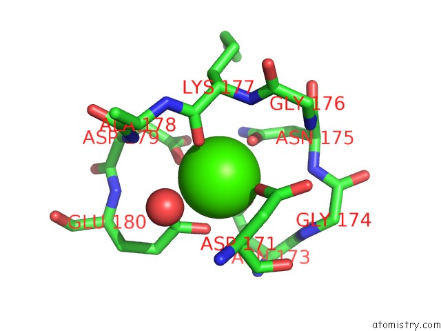
Mono view
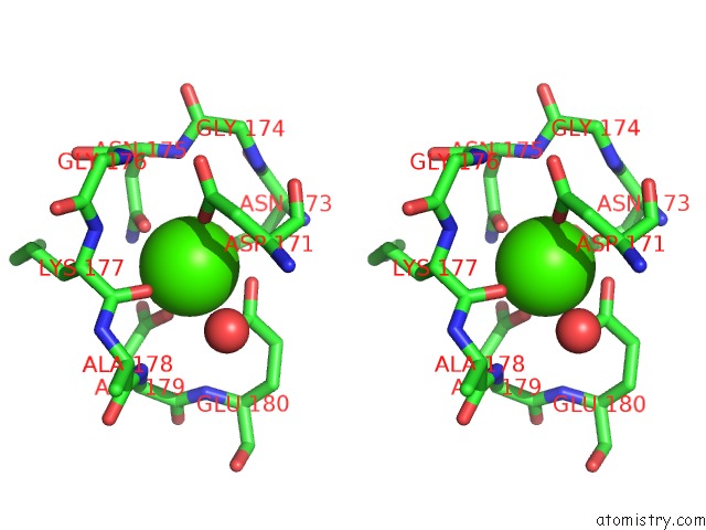
Stereo pair view
Mono view
Stereo pair view
A full contact list of Calcium with other atoms in the Ca binding
site number 1 of Crystal Structure of Sphingomonas Sp. A1 Alginate-Binding Ptotein ALGQ1 in Complex with Unsaturated Triguluronate within 5.0Å range:
|
Reference:
Y.Nishitani,
Y.Maruyama,
T.Itoh,
B.Mikami,
W.Hashimoto,
K.Murata.
Recognition of Heteropolysaccharide Alginate By Periplasmic Solute-Binding Proteins of A Bacterial Abc Transporter Biochemistry V. 51 3622 2012.
ISSN: ISSN 0006-2960
PubMed: 22486720
DOI: 10.1021/BI300194F
Page generated: Tue Jul 8 17:39:00 2025
ISSN: ISSN 0006-2960
PubMed: 22486720
DOI: 10.1021/BI300194F
Last articles
Mg in 8ZWBMg in 8ZWQ
Mg in 8ZUT
Mg in 8ZVC
Mg in 8ZUS
Mg in 8ZTZ
Mg in 8ZUQ
Mg in 8ZK2
Mg in 8ZUP
Mg in 8ZTF