Calcium »
PDB 3whi-3ws5 »
3wqr »
Calcium in PDB 3wqr: Crystal Structure of Pfdxr Complexed with Inhibitor-12
Enzymatic activity of Crystal Structure of Pfdxr Complexed with Inhibitor-12
All present enzymatic activity of Crystal Structure of Pfdxr Complexed with Inhibitor-12:
1.1.1.267;
1.1.1.267;
Protein crystallography data
The structure of Crystal Structure of Pfdxr Complexed with Inhibitor-12, PDB code: 3wqr
was solved by
N.Tanaka,
T.Umeda,
with X-Ray Crystallography technique. A brief refinement statistics is given in the table below:
| Resolution Low / High (Å) | 50.00 / 1.97 |
| Space group | P 1 21 1 |
| Cell size a, b, c (Å), α, β, γ (°) | 51.081, 76.551, 104.095, 90.00, 90.68, 90.00 |
| R / Rfree (%) | 18 / 23 |
Other elements in 3wqr:
The structure of Crystal Structure of Pfdxr Complexed with Inhibitor-12 also contains other interesting chemical elements:
| Magnesium | (Mg) | 2 atoms |
Calcium Binding Sites:
The binding sites of Calcium atom in the Crystal Structure of Pfdxr Complexed with Inhibitor-12
(pdb code 3wqr). This binding sites where shown within
5.0 Angstroms radius around Calcium atom.
In total 2 binding sites of Calcium where determined in the Crystal Structure of Pfdxr Complexed with Inhibitor-12, PDB code: 3wqr:
Jump to Calcium binding site number: 1; 2;
In total 2 binding sites of Calcium where determined in the Crystal Structure of Pfdxr Complexed with Inhibitor-12, PDB code: 3wqr:
Jump to Calcium binding site number: 1; 2;
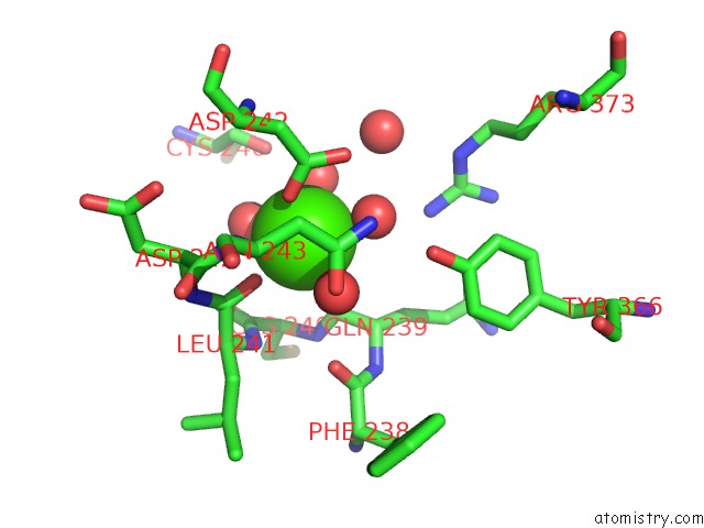
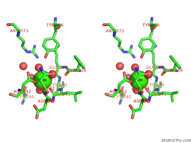
Calcium binding site 1 out of 2 in 3wqr
Go back to
Calcium binding site 1 out
of 2 in the Crystal Structure of Pfdxr Complexed with Inhibitor-12
Mono view
Stereo pair view
Mono view
Stereo pair view
A full contact list of Calcium with other atoms in the Ca binding
site number 1 of Crystal Structure of Pfdxr Complexed with Inhibitor-12 within 5.0Å range:
|
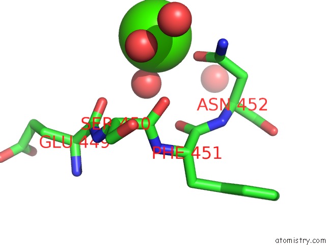
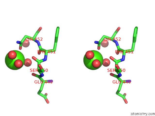
Calcium binding site 2 out of 2 in 3wqr
Go back to
Calcium binding site 2 out
of 2 in the Crystal Structure of Pfdxr Complexed with Inhibitor-12
Mono view
Stereo pair view
Mono view
Stereo pair view
A full contact list of Calcium with other atoms in the Ca binding
site number 2 of Crystal Structure of Pfdxr Complexed with Inhibitor-12 within 5.0Å range:
|
Reference:
S.Konzuch,
T.Umeda,
J.Held,
S.Hahn,
K.Brucher,
C.Lienau,
C.T.Behrendt,
T.Grawert,
A.Bacher,
B.Illarionov,
M.Fischer,
B.Mordmuller,
N.Tanaka,
T.Kurz.
Binding Modes of Reverse Fosmidomycin Analogs Toward the Antimalarial Target Ispc. J.Med.Chem. V. 57 8827 2014.
ISSN: ISSN 0022-2623
PubMed: 25254502
DOI: 10.1021/JM500850Y
Page generated: Tue Jul 8 18:01:39 2025
ISSN: ISSN 0022-2623
PubMed: 25254502
DOI: 10.1021/JM500850Y
Last articles
Mg in 7D8QMg in 7D8L
Mg in 7D7Z
Mg in 7D82
Mg in 7D8G
Mg in 7D86
Mg in 7D81
Mg in 7D7Y
Mg in 7D7X
Mg in 7D7W