Calcium »
PDB 3wt3-3zq4 »
3zhq »
Calcium in PDB 3zhq: Crystal Structure of the H747A Mutant of the Suca Domain of Mycobacterium Smegmatis Kgd
Enzymatic activity of Crystal Structure of the H747A Mutant of the Suca Domain of Mycobacterium Smegmatis Kgd
All present enzymatic activity of Crystal Structure of the H747A Mutant of the Suca Domain of Mycobacterium Smegmatis Kgd:
1.2.4.2; 2.2.1.5; 2.3.1.61; 4.1.1.71;
1.2.4.2; 2.2.1.5; 2.3.1.61; 4.1.1.71;
Protein crystallography data
The structure of Crystal Structure of the H747A Mutant of the Suca Domain of Mycobacterium Smegmatis Kgd, PDB code: 3zhq
was solved by
T.Wagner,
N.Barilone,
M.Bellinzoni,
P.M.Alzari,
with X-Ray Crystallography technique. A brief refinement statistics is given in the table below:
| Resolution Low / High (Å) | 48.97 / 2.50 |
| Space group | P 1 |
| Cell size a, b, c (Å), α, β, γ (°) | 80.675, 83.737, 159.577, 99.86, 98.95, 100.44 |
| R / Rfree (%) | 19.37 / 22.34 |
Other elements in 3zhq:
The structure of Crystal Structure of the H747A Mutant of the Suca Domain of Mycobacterium Smegmatis Kgd also contains other interesting chemical elements:
| Magnesium | (Mg) | 4 atoms |
Calcium Binding Sites:
The binding sites of Calcium atom in the Crystal Structure of the H747A Mutant of the Suca Domain of Mycobacterium Smegmatis Kgd
(pdb code 3zhq). This binding sites where shown within
5.0 Angstroms radius around Calcium atom.
In total 4 binding sites of Calcium where determined in the Crystal Structure of the H747A Mutant of the Suca Domain of Mycobacterium Smegmatis Kgd, PDB code: 3zhq:
Jump to Calcium binding site number: 1; 2; 3; 4;
In total 4 binding sites of Calcium where determined in the Crystal Structure of the H747A Mutant of the Suca Domain of Mycobacterium Smegmatis Kgd, PDB code: 3zhq:
Jump to Calcium binding site number: 1; 2; 3; 4;
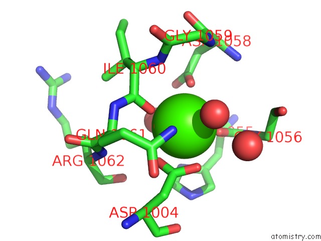
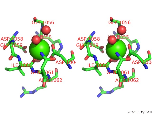
Calcium binding site 1 out of 4 in 3zhq
Go back to
Calcium binding site 1 out
of 4 in the Crystal Structure of the H747A Mutant of the Suca Domain of Mycobacterium Smegmatis Kgd
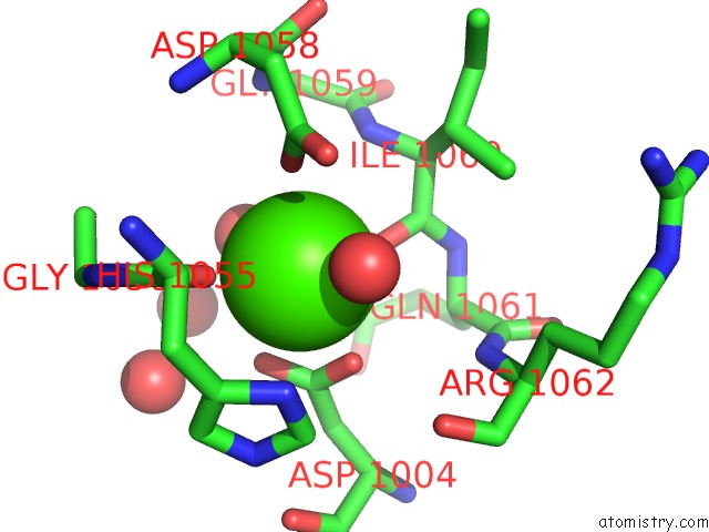
Mono view
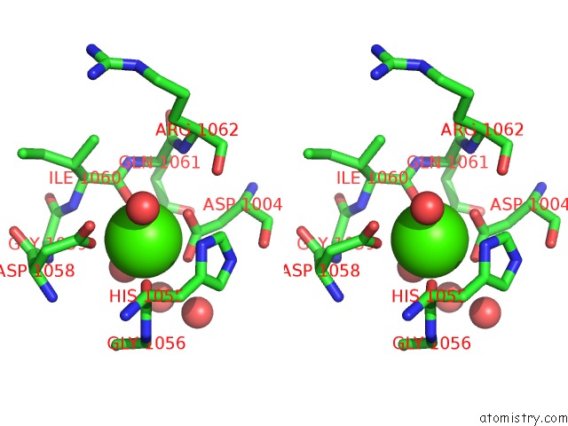
Stereo pair view
Mono view
Stereo pair view
A full contact list of Calcium with other atoms in the Ca binding
site number 1 of Crystal Structure of the H747A Mutant of the Suca Domain of Mycobacterium Smegmatis Kgd within 5.0Å range:
|
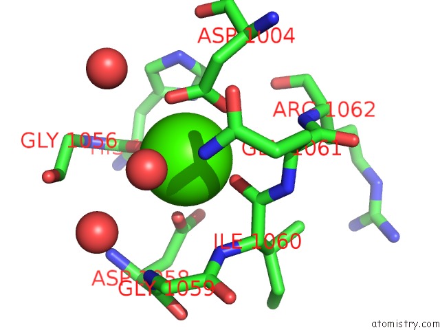
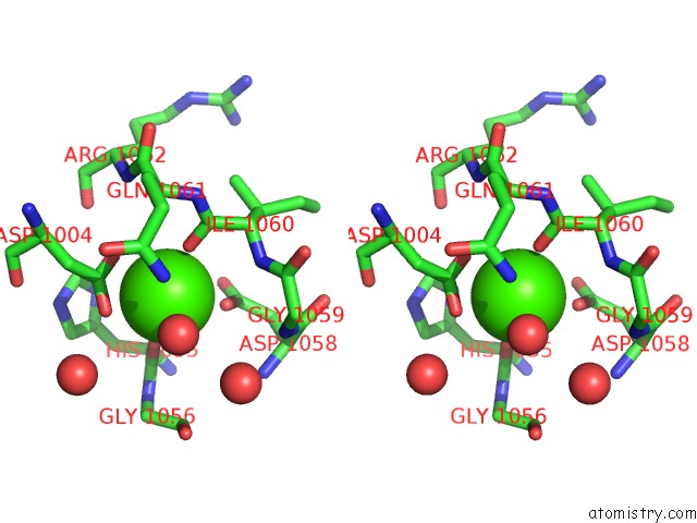
Calcium binding site 2 out of 4 in 3zhq
Go back to
Calcium binding site 2 out
of 4 in the Crystal Structure of the H747A Mutant of the Suca Domain of Mycobacterium Smegmatis Kgd
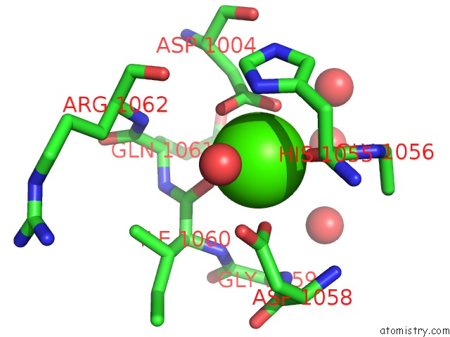
Mono view
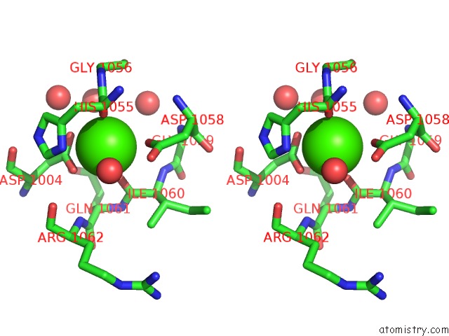
Stereo pair view
Mono view
Stereo pair view
A full contact list of Calcium with other atoms in the Ca binding
site number 2 of Crystal Structure of the H747A Mutant of the Suca Domain of Mycobacterium Smegmatis Kgd within 5.0Å range:
|
Calcium binding site 3 out of 4 in 3zhq
Go back to
Calcium binding site 3 out
of 4 in the Crystal Structure of the H747A Mutant of the Suca Domain of Mycobacterium Smegmatis Kgd
Mono view
Stereo pair view
Mono view
Stereo pair view
A full contact list of Calcium with other atoms in the Ca binding
site number 3 of Crystal Structure of the H747A Mutant of the Suca Domain of Mycobacterium Smegmatis Kgd within 5.0Å range:
|
Calcium binding site 4 out of 4 in 3zhq
Go back to
Calcium binding site 4 out
of 4 in the Crystal Structure of the H747A Mutant of the Suca Domain of Mycobacterium Smegmatis Kgd
Mono view
Stereo pair view
Mono view
Stereo pair view
A full contact list of Calcium with other atoms in the Ca binding
site number 4 of Crystal Structure of the H747A Mutant of the Suca Domain of Mycobacterium Smegmatis Kgd within 5.0Å range:
|
Reference:
T.Wagner,
N.Barilone,
P.M.Alzari,
M.Bellinzoni.
A Dual Conformation of the Post-Decarboxylation Intermediate Is Associated with Distinct Enzyme States in Mycobacterial Alpha-Ketoglutarate Decarboxylase (Kgd). Biochem.J. V. 457 425 2014.
ISSN: ISSN 0264-6021
PubMed: 24171907
DOI: 10.1042/BJ20131142
Page generated: Tue Jul 8 18:13:42 2025
ISSN: ISSN 0264-6021
PubMed: 24171907
DOI: 10.1042/BJ20131142
Last articles
Mg in 4GMJMg in 4GNK
Mg in 4GNI
Mg in 4GN0
Mg in 4GMX
Mg in 4GME
Mg in 4GKM
Mg in 4GKR
Mg in 4GHL
Mg in 4GIU