Calcium »
PDB 4ae2-4aqe »
4aj3 »
Calcium in PDB 4aj3: 3D Structure of E. Coli Isocitrate Dehydrogenase in Complex with Isocitrate, Calcium(II) and Nadp - the Pseudo-Michaelis Complex
Enzymatic activity of 3D Structure of E. Coli Isocitrate Dehydrogenase in Complex with Isocitrate, Calcium(II) and Nadp - the Pseudo-Michaelis Complex
All present enzymatic activity of 3D Structure of E. Coli Isocitrate Dehydrogenase in Complex with Isocitrate, Calcium(II) and Nadp - the Pseudo-Michaelis Complex:
1.1.1.42;
1.1.1.42;
Protein crystallography data
The structure of 3D Structure of E. Coli Isocitrate Dehydrogenase in Complex with Isocitrate, Calcium(II) and Nadp - the Pseudo-Michaelis Complex, PDB code: 4aj3
was solved by
S.Goncalves,
S.P.Miller,
M.A.Carrondo,
A.M.Dean,
P.M.Matias,
with X-Ray Crystallography technique. A brief refinement statistics is given in the table below:
| Resolution Low / High (Å) | 44.81 / 1.90 |
| Space group | P 43 21 2 |
| Cell size a, b, c (Å), α, β, γ (°) | 105.297, 105.297, 145.723, 90.00, 90.00, 90.00 |
| R / Rfree (%) | 18.9 / 22.4 |
Calcium Binding Sites:
The binding sites of Calcium atom in the 3D Structure of E. Coli Isocitrate Dehydrogenase in Complex with Isocitrate, Calcium(II) and Nadp - the Pseudo-Michaelis Complex
(pdb code 4aj3). This binding sites where shown within
5.0 Angstroms radius around Calcium atom.
In total only one binding site of Calcium was determined in the 3D Structure of E. Coli Isocitrate Dehydrogenase in Complex with Isocitrate, Calcium(II) and Nadp - the Pseudo-Michaelis Complex, PDB code: 4aj3:
In total only one binding site of Calcium was determined in the 3D Structure of E. Coli Isocitrate Dehydrogenase in Complex with Isocitrate, Calcium(II) and Nadp - the Pseudo-Michaelis Complex, PDB code: 4aj3:
Calcium binding site 1 out of 1 in 4aj3
Go back to
Calcium binding site 1 out
of 1 in the 3D Structure of E. Coli Isocitrate Dehydrogenase in Complex with Isocitrate, Calcium(II) and Nadp - the Pseudo-Michaelis Complex
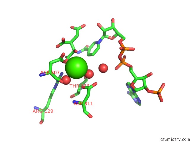
Mono view
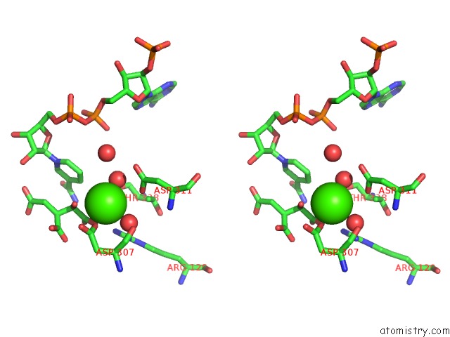
Stereo pair view
Mono view
Stereo pair view
A full contact list of Calcium with other atoms in the Ca binding
site number 1 of 3D Structure of E. Coli Isocitrate Dehydrogenase in Complex with Isocitrate, Calcium(II) and Nadp - the Pseudo-Michaelis Complex within 5.0Å range:
|
Reference:
S.Goncalves,
S.P.Miller,
M.A.Carrondo,
A.M.Dean,
P.M.Matias.
Induced Fit and the Catalytic Mechanism of Isocitrate Dehydrogenase. Biochemistry V. 51 7098 2012.
ISSN: ISSN 0006-2960
PubMed: 22891681
DOI: 10.1021/BI300483W
Page generated: Tue Jul 8 18:31:12 2025
ISSN: ISSN 0006-2960
PubMed: 22891681
DOI: 10.1021/BI300483W
Last articles
Mg in 6TR4Mg in 6TR3
Mg in 6TMF
Mg in 6TQO
Mg in 6TQN
Mg in 6TQF
Mg in 6TQE
Mg in 6TQB
Mg in 6TQA
Mg in 6TPS