Calcium »
PDB 4bw8-4cg0 »
4cg0 »
Calcium in PDB 4cg0: Savinase Crystal Structures For Combined Single Crystal Diffraction and Powder Diffraction Analysis
Enzymatic activity of Savinase Crystal Structures For Combined Single Crystal Diffraction and Powder Diffraction Analysis
All present enzymatic activity of Savinase Crystal Structures For Combined Single Crystal Diffraction and Powder Diffraction Analysis:
3.4.21.62;
3.4.21.62;
Protein crystallography data
The structure of Savinase Crystal Structures For Combined Single Crystal Diffraction and Powder Diffraction Analysis, PDB code: 4cg0
was solved by
C.G.Frankaer,
O.V.Moroz,
J.P.Turkenburg,
S.I.Aspmo,
M.Thymark,
E.P.Friis,
K.Stahla,
J.E.Nielsen,
K.S.Wilson,
P.Harris,
with X-Ray Crystallography technique. A brief refinement statistics is given in the table below:
| Resolution Low / High (Å) | 44.80 / 1.36 |
| Space group | P 21 21 21 |
| Cell size a, b, c (Å), α, β, γ (°) | 53.040, 57.550, 71.370, 90.00, 90.00, 90.00 |
| R / Rfree (%) | 10.908 / 15.413 |
Other elements in 4cg0:
The structure of Savinase Crystal Structures For Combined Single Crystal Diffraction and Powder Diffraction Analysis also contains other interesting chemical elements:
| Sodium | (Na) | 1 atom |
Calcium Binding Sites:
The binding sites of Calcium atom in the Savinase Crystal Structures For Combined Single Crystal Diffraction and Powder Diffraction Analysis
(pdb code 4cg0). This binding sites where shown within
5.0 Angstroms radius around Calcium atom.
In total only one binding site of Calcium was determined in the Savinase Crystal Structures For Combined Single Crystal Diffraction and Powder Diffraction Analysis, PDB code: 4cg0:
In total only one binding site of Calcium was determined in the Savinase Crystal Structures For Combined Single Crystal Diffraction and Powder Diffraction Analysis, PDB code: 4cg0:
Calcium binding site 1 out of 1 in 4cg0
Go back to
Calcium binding site 1 out
of 1 in the Savinase Crystal Structures For Combined Single Crystal Diffraction and Powder Diffraction Analysis
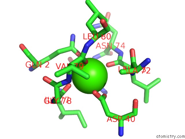
Mono view
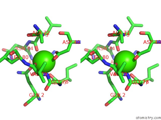
Stereo pair view
Mono view
Stereo pair view
A full contact list of Calcium with other atoms in the Ca binding
site number 1 of Savinase Crystal Structures For Combined Single Crystal Diffraction and Powder Diffraction Analysis within 5.0Å range:
|
Reference:
C.G.Frankaer,
O.V.Moroz,
J.P.Turkenburg,
S.I.Aspmo,
M.Thymark,
E.P.Friis,
K.Stahl,
J.E.Nielsen,
K.S.Wilson,
P.Harris.
Analysis of An Industrial Production Suspension of Bacillus Lentus Subtilisin Crystals By Powder Diffraction: A Powerful Quality-Control Tool. Acta Crystallogr.,Sect.D V. 70 1115 2014.
ISSN: ISSN 0907-4449
PubMed: 24699655
DOI: 10.1107/S1399004714001497
Page generated: Tue Jul 8 19:12:54 2025
ISSN: ISSN 0907-4449
PubMed: 24699655
DOI: 10.1107/S1399004714001497
Last articles
Mn in 9LJUMn in 9LJW
Mn in 9LJS
Mn in 9LJR
Mn in 9LJT
Mn in 9LJV
Mg in 9UA2
Mg in 9R96
Mg in 9VM1
Mg in 9P01